When a dog needs diagnostic testing, the first step is obtaining a high-quality blood sample. In clinical practice, the two primary sites for venipuncture are the front leg and the neck. But which one is best?
Whether you are a veterinary student perfecting your technique or a pet owner curious about the process, understanding the nuances between a dog cephalic vs. jugular vein blood draws is essential for patient care.
For many veterinarians, the dog jugular vein blood draw is the preferred method, especially when a large volume of blood is required for comprehensive health panels.
Why the Dog Jugular Vein Blood Draw is Often the Gold Standard
- Speed and Efficiency: The jugular is a large-diameter vessel, allowing for rapid collection—crucial for stressed or wiggly patients.
- Superior Sample Quality: A fast, steady flow reduces the risk of hemolysis (the rupturing of red blood cells), ensuring that lab results for chemistry and electrolytes are 100% accurate.
- Saving Peripheral Veins: By using the neck for blood samples, the veins in the legs remain healthy and undamaged in case the dog requires an IV catheter for surgery or fluid therapy later.
When is a Dog Cephalic Vein Blood Draw Used?
The cephalic vein runs along the top of the front leg. While it is very accessible, it is a much smaller vessel. Veterinarians typically choose the dog cephalic vein blood draw when:
- Only a tiny amount of blood is needed (like a quick heartworm snap test).
- The dog has a neck injury or is extremely sensitive about their throat area.
- The dog is positioned in a way that makes the front legs more reachable than the neck.
Comparison: Advantages and Challenges
Choosing the right site is a balance of clinical needs and the patient’s specific condition. Here is a breakdown of the pros and cons:
1. Dog Jugular Vein (The Neck)
- Advantages: * Ideal for collecting large volumes (5ml+).
- Less likely to clot during collection.
- Generally less painful for the dog as the skin on the neck is less sensitive than the paws.
- Challenges: * Requires precise restraint to keep the neck extended and head steady.
- Can be difficult to palpate (feel) in obese dogs or breeds with thick skin folds like Bulldogs.
2. Dog Cephalic Vein (The Front Leg)
- Advantages: * Highly accessible and visually easy to locate.
- Perfect for quick, small-volume tests (like Heartworm or Glucose checks).
- Easier to apply pressure to stop bleeding afterward.
- Challenges: * Vein Collapse: Because it is a smaller vessel, pulling the syringe plunger too hard can cause the vein to collapse, halting the draw.
- Movement Issues: Dogs often reflexively pull their paws away, increasing the risk of “blowing” the vein.
| Feature | Dog Jugular Vein | Dog Cephalic Vein |
| Vessel Size | Large | Small to Medium |
| Collection Speed | Very Fast | Moderate to Slow |
| Common Needle Gauge | 21G – 22G | 23G – 25G |
| Best For | Full Senior Panels, ER Work | Quick Tests, Small Breeds |
| Success Rate | High (with skilled restraint) | High (for small volumes) |
Special Focus: Why the Jugular Vein is Often Better for Large Breeds
For owners of larger breeds—like Golden Retrievers, German Shepherds, or Great Danes—seeing a vet reach for the neck instead of the leg can be surprising. However, the dog jugular vein blood draw is often the safest and most humane choice for big dogs for several clinical reasons:
1. Thicker Skin and Denser Fur on Limbs
Large breeds often have much tougher skin and denser undercoats on their legs. A dog cephalic vein blood draw on a thick-skinned breed requires more force to penetrate, which can increase the “pinch” sensation for the dog. The skin over the jugular vein is generally thinner and more elastic, allowing the needle to glide in with much less resistance.
2. Reduced Risk of “Needle Shyness”
Large dogs are powerful. If a big dog decides to jerk its leg during a cephalic draw, it can lead to a “blown vein,” a bent needle, or a painful hematoma. Because the neck is a more stable central axis, a professional holder can keep a large dog much calmer and steadier, making the dog jugular vein blood draw a quicker “one-and-done” experience.
3. Preserving the “Life-Line”
In large breed dogs, health issues like bloat (GDV) or joint surgeries are more common. These conditions require emergency or long-term IV fluid therapy. By consistently choosing the jugular for routine blood work, vets preserve the cephalic veins (front legs) as pristine sites for emergency catheters.
4. Avoiding “Pump-Action” Hemolysis
Large dogs have a high blood volume. Trying to pull a large sample through the small cephalic vein often creates a vacuum struggle, leading to hemolysis. The jugular provides a high-pressure, high-volume flow that fills the syringe effortlessly, ensuring the lab gets a perfect sample on the first try.
Mastering the Hold: Dog Restraint Techniques
A successful blood draw is 50% technique and 50% restraint. If the dog moves, even the most skilled vet can “blow” a vein. Here is how to properly secure a dog for each method:
1. Restraining for a Dog Cephalic Vein Blood Draw
The goal here is to “roll” the vein to make it visible and prevent the leg from pulling back.
- The Position: The dog can be sitting or lying down (sternal recumbency).
- The Grip: Stand on the opposite side of the leg being used. Reach over the dog’s back, cradling their head under your arm to prevent biting or sudden lunges.
- The “Roll”: Place your thumb on the medial (inner) side of the dog’s elbow and wrap your fingers around the back. Push your thumb toward the outside (lateral) of the leg. This “rolls” the cephalic vein to the top of the limb, making it a stable target.
- The Lock: Use your other hand to hold the dog’s paw from behind the “wrist” (carpus) to keep the leg extended.
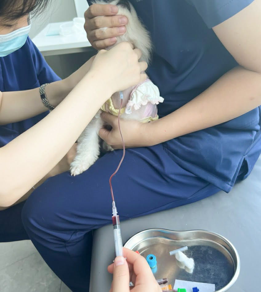
2. Restraining for a Dog Jugular Vein Blood Draw
This requires the dog to be steady and the neck to be elongated to expose the jugular furrow.
- The Position: Most dogs are best restrained in a sitting position or at the edge of the exam table.
- The “Nose to the Sky”: Place one hand under the dog’s muzzle and gently lift their head toward the ceiling. Do not over-extend, as this can collapse the vein. The goal is a straight line from the jaw to the chest.
- Body Control: Use your legs or your other arm to hug the dog’s body against yours. This prevents them from scooting backward or shifting their weight.
- The Target: The person drawing the blood will “occlude” (apply pressure) at the base of the neck, and the vein will pop up like a small rope along the side of the windpipe.
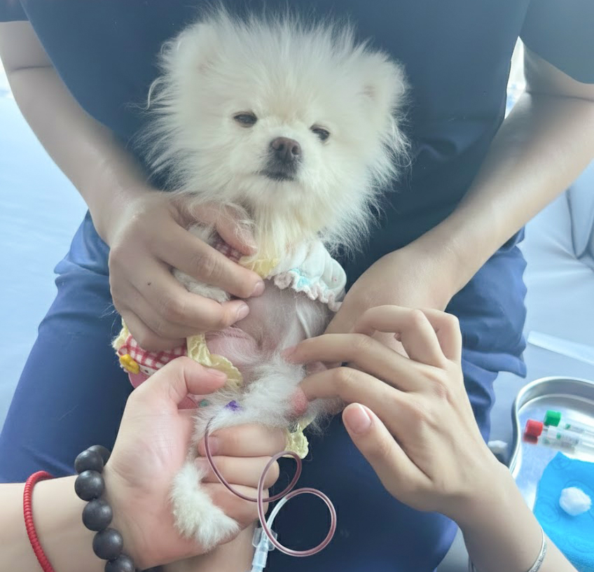
Safety Tips for Restrainers
- Watch the Mouth: Even the sweetest dog may nip when feeling a “pinch.” Always maintain control of the head.
- Less is More: Use the minimum amount of force necessary. Excessive squeezing can make a dog panic, causing them to struggle more.
- The “Lick Mat” Trick: For a jugular draw, having a second assistant hold a lick mat with peanut butter can keep the dog’s head perfectly still while they focus on the treat.
Useful tools
Medtacedu offers dog cephalic blood draw model for veterinary practice. Through repeated practice, students could develop muscle memory before practice on real patients.
Meanwhile, MedDimension provides dog jugular access model for jugular blood draw training.
Key Takeaways: What You Need to Know
- Size Matters: The dog jugular vein blood draw is the preferred method for large-volume samples (like senior or emergency panels) due to its high flow rate and superior sample quality.
- Preserving the “Life-Line”: Using the jugular for routine blood work leaves the cephalic (leg) veins healthy and available for IV catheters should the dog ever need surgery or emergency fluids.
- Large Breed Advantage: For big dogs, the jugular is often less painful than the leg because the skin is thinner and the dog is easier to keep steady, reducing the risk of bruising.
- Restraint is 50% of Success: Proper technique—like “rolling” the cephalic vein or the “nose-to-the-sky” position for the jugular—is essential to prevent hematomas and ensure a “first-stick” success.
- Sample Integrity: Larger veins and correct needle gauges (like 21G or 22G) significantly reduce the risk of hemolysis, ensuring your lab results are accurate the first time.