The external jugular vein is a useful blood-collection site in dogs because it is large, has brisk flow, and can provide an adequate diagnostic sample without using a peripheral vein that may later be needed for an IV catheter. Those advantages only apply when patient selection, restraint, vessel occlusion, needle control, and post-draw pressure are all handled well.
This guide focuses only on dog jugular vein phlebotomy. For a comparison of the jugular, cephalic, and saphenous veins, see our broader canine venipuncture and blood collection guide.
Scope: This page covers canine jugular blood collection. It does not teach peripheral IV catheter placement, SC/IM medication injection, or feline IV access. See canine IV catheterization, dog SC vs IM injections, and the Veterinary Cat IV Practice Kit for those separate skills.
Important: Jugular venipuncture is a clinical procedure for trained veterinary personnel and supervised learners. It is not an at-home blood-draw technique. Follow the veterinarian’s instructions, the laboratory’s specimen requirements, and your practice’s restraint and sharps protocols.
When Is the Jugular Vein a Good Choice?
The jugular is often considered when the team needs a moderate or larger sample, rapid blood flow, or preservation of the cephalic veins for possible catheter placement. A clean jugular draw can also reduce the prolonged aspiration and repeated manipulation that damage samples from a poorly filling peripheral vein.
It is not automatically the best site for every patient. Site selection should account for the dog’s behavior, body position, neck anatomy, cardiovascular status, required sample volume, planned tests, and bleeding risk.
Avoid or reconsider jugular venipuncture when there is a suspected coagulation disorder, clinically important thrombocytopenia, anticoagulant toxicity, disseminated intravascular coagulation, or another serious hemostatic defect. A cervical hematoma can threaten the airway, so Cornell’s Comparative Coagulation Laboratory recommends peripheral veins for patients with severe hemostatic defects. Neck trauma, infection over the intended site, an existing jugular catheter, or a patient that cannot safely tolerate head and neck positioning may also require a different plan.
Canine Jugular Anatomy and Surface Landmarks
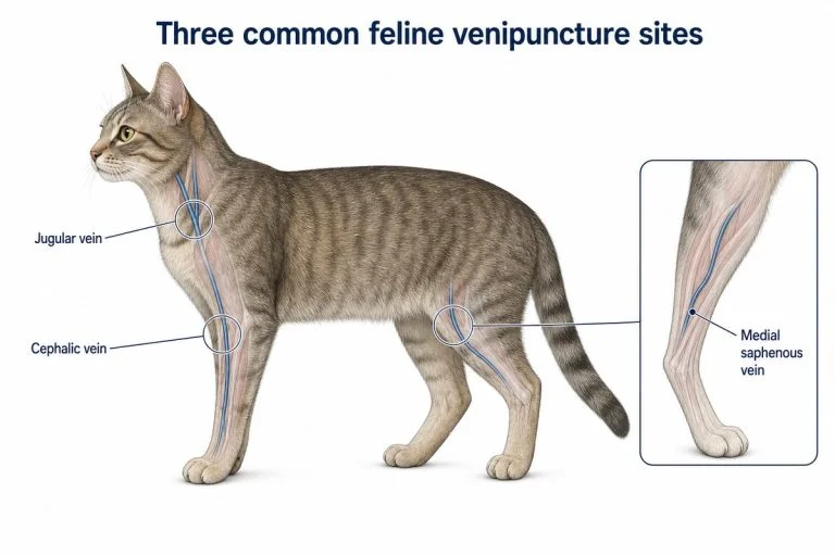
The external jugular vein runs in the jugular groove on each side of the neck, from near the angle of the mandible toward the thoracic inlet. It is lateral to the trachea, not directly over it.
The vein is usually occluded near the thoracic inlet so it fills cranial to the point of pressure. The University of Minnesota clinical skills guide recommends using the non-dominant thumb at the thoracic inlet, then watching and palpating the jugular area as the vessel distends. Virginia Tech’s venipuncture SOP describes the middle third of the external jugular as the usual puncture region.
Do not rely on coat pattern or sight alone. In many dogs, the vein is easier to palpate than to see. Correct occlusion should produce a vessel that fills when pressure is applied and softens when pressure is released.
Positioning and Restraint
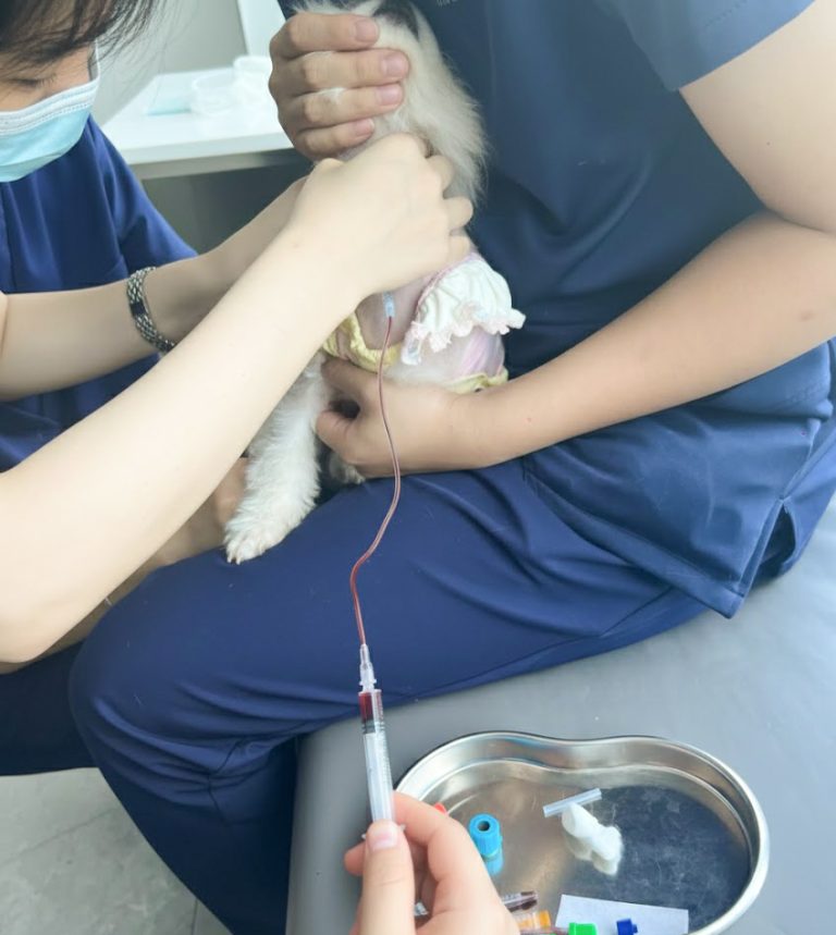
A successful canine jugular blood draw is a team procedure. The restrainer controls the body and head while the person drawing blood locates, occludes, enters, and samples the vein. Both people should agree on the sequence and on the words used before the needle is uncapped.
Sitting or standing
Many dogs can remain sitting or standing on a non-slip surface. A large dog may be positioned with its back near a wall or stable table so it cannot back away. The restrainer supports the dog from the side, gently elevates the chin, and keeps the neck aligned without forcing the muzzle closed.
Virginia Tech describes the neck as ideally near a right angle to the thoracic spine. This exposes the jugular grooves, but the exact head height should be adjusted to the individual dog. Excessive extension can make the patient uncomfortable and may not improve venous filling.
Lateral recumbency
Lateral positioning can be useful for a small dog, a patient that is more comfortable lying down, or a team trained in a lateral jugular technique. Support the body and keep the neck in a neutral, sustainable position. The chosen position should reduce movement without escalating fear or compromising breathing.
Low-stress handling principles
Use the least restraint that safely controls the patient. Avoid leaning over the dog or clamping the muzzle tightly. Keep one side visually open when possible, use calm team communication, and pause if fear, struggling, respiratory effort, or bite risk increases. A different position, a different site, pre-visit medication, local anesthetic, or chemical restraint may be safer than overpowering the patient.
Equipment and Sample Planning
Prepare every item before positioning the dog:
- The test request and required blood volume
- Correct collection tubes and labels
- An appropriately sized sterile needle and syringe, butterfly set, or evacuated collection system
- Gauze and an approved skin antiseptic
- Clippers if hair removal is clinically indicated or required by protocol
- A sharps container within reach
- Any practice-specific personal protective equipment
Needle and syringe selection depends on patient size, vein size, test requirements, and collection method. A very small needle is not always gentler: narrow lumens and strong syringe suction can increase red-cell trauma. Conversely, a large evacuated tube can collapse a small or poorly filled vein. Choose equipment that allows a prompt sample with controlled negative pressure.
Know the tubes, fill requirements, and order specified by the receiving laboratory. This is especially important for coagulation samples, where the blood-to-citrate ratio must be correct.
Step-by-Step Dog Jugular Blood Draw
1. Confirm the patient and collection plan
Verify the dog’s identity, requested tests, fasting or timing instructions, required volume, and any history that changes bleeding or restraint risk. Check for anticoagulant use, thrombocytopenia, neck lesions, respiratory compromise, and previous difficult draws.
2. Brief the team and position the dog
Agree on who controls the head, who controls the body, and what verbal cue will signal needle entry and withdrawal. Place the dog on a stable, non-slip surface. Align and gently elevate the head enough to expose the selected jugular groove.
3. Identify the jugular groove
Palpate lateral to the trachea from the angle of the mandible toward the thoracic inlet. Part or wet the coat if appropriate. Clip hair when visualization, asepsis, or practice protocol requires it.
4. Occlude and confirm the vessel
Use the non-dominant thumb to compress the vein near the thoracic inlet. Palpate the vessel as it fills. Releasing and reapplying pressure should make the structure soften and refill; this helps distinguish the vein from the trachea, muscle, or a fixed cord-like structure.
Apply light skin tension to stabilize the vessel. If the vein does not fill, reset the head, neck, thumb position, and body alignment before reaching for the needle.
5. Prepare the site
Clean the intended puncture area according to practice protocol and let the antiseptic work as directed. Aseptic preparation is essential when collecting blood for culture. Do not repalpate a prepared culture site with an unsterile finger.
6. Enter the vein with a controlled, shallow approach
Hold the needle bevel up and direct it toward the head. The approach is generally shallow; University of Minnesota teaching material describes approximately 15 to 20 degrees, while other veterinary SOPs use an angle around 25 degrees depending on anatomy and equipment.
Enter the middle third of the distended jugular with one deliberate motion. Avoid sweeping or “fishing” under the skin. Stabilizing the syringe against the non-dominant hand can reduce unwanted movement.
7. Confirm placement and collect smoothly
Look for blood at the hub or in the syringe. Once the bevel is within the lumen, aspirate with steady, modest negative pressure. Excessive or fluctuating suction can collapse the vein and contribute to hemolysis.
Keep the needle still while the syringe plunger moves. If flow stops, do not immediately probe in multiple directions. First reduce suction and reassess whether the vein is still occluded, the dog has moved, or the bevel is only partly within the lumen.
8. Release occlusion before withdrawing
When the required volume has been collected, release pressure at the thoracic inlet and relax syringe suction. Tell the restrainer that the needle is coming out. This reduces pressure within the vein before withdrawal.
9. Withdraw and apply immediate pressure
Remove the needle in one smooth motion while the restrainer places folded gauze over the site. Apply direct, gentle pressure without repeatedly lifting the gauze to check. University of Minnesota teaching guidance uses at least 30 seconds for routine patients; continue longer when the site is still oozing or the clinician directs it.
Confirm hemostasis and check for swelling before releasing the dog.
10. Transfer and handle the sample correctly
If using a syringe, remove the needle before transferring blood when the tube system allows; forcing blood through a narrow needle can rupture cells. Fill additive tubes to the laboratory’s required level and mix by gentle inversion, not vigorous shaking. Label the specimen at the patient side and dispose of the needle immediately in the sharps container.
Protecting Sample Quality
The blood draw is not successful if the sample cannot answer the clinical question. Common pre-analytical problems include hemolysis, clotting, platelet clumping, and an incorrect blood-to-additive ratio.
To reduce hemolysis:
- Use clean, dry collection equipment
- Avoid repeated punctures and traumatic needle movement
- Use controlled rather than forceful or fluctuating aspiration
- Do not force blood through the attached needle into a tube
- Mix additive tubes gently instead of shaking them
To reduce clotting or invalid fill ratios:
- Have the correct tubes open and ready before the draw
- Transfer promptly
- Fill tubes to the level required by the laboratory
- Mix anticoagulant tubes immediately using the laboratory’s inversion guidance
- Redraw a coagulation specimen if the citrate ratio or sample integrity is unacceptable
Common Technique Errors
Occluding over the trachea
The external jugular is lateral to the trachea. Pressure in the wrong place will not distend the vessel and may make the dog resist positioning.
Extending the head too far
More extension is not always better. Overextension can increase discomfort, particularly in a senior dog or a patient with neck pain, and may pull soft tissue tight enough to make the vessel harder to stabilize.
Entering too steeply
A steep approach can pass through the front and back walls. Reset to a shallower angle that matches the vessel’s depth.
Pulling the plunger too hard
Strong suction can collapse the vein against the bevel or damage the sample. Use a syringe size and collection system that allow controlled aspiration.
Withdrawing while the vein is still occluded
Release the thoracic-inlet pressure and syringe suction before removing the needle, then apply gauze immediately. This sequence helps limit bleeding and hematoma formation.
Repeatedly redirecting beneath the skin
Blind probing traumatizes tissue and the vessel. Stop, withdraw, apply pressure, and reassess. A second trained person or a different site is better than accumulating damage in one area.
When to Stop and Choose Another Plan
Stop the attempt when the dog cannot be restrained without escalating fear or compromising breathing, when a hematoma begins to form, when several controlled attempts have failed, or when new information suggests an unsafe bleeding risk. Apply pressure, reassess the patient, and let the responsible clinician choose a different operator, vein, collection method, or stabilization plan.
Conclusion
Reliable dog jugular vein phlebotomy depends on a sequence, not a single needle movement: select an appropriate patient, expose and confirm the jugular groove, coordinate low-stress restraint, use controlled aspiration, release occlusion before withdrawal, and maintain pressure until hemostasis is secure. When the vein will not fill, flow stops, swelling develops, or bleeding risk changes, stopping and reassessing is safer than accumulating attempts. For failure patterns, complications, and special-patient adaptations, see Difficult Canine Jugular Venipuncture.
Frequently Asked Questions
Can either jugular vein be used for a dog’s blood draw?
Either external jugular may be suitable. The veterinary team chooses the side with the clearest landmark, safest positioning, and no local swelling, skin lesion, previous trauma, or vascular device that makes access inappropriate. If one side has already been punctured or bruised, the clinician should reassess before using that area again.
Does a dog need sedation for jugular venipuncture?
Most calm, cooperative dogs do not need sedation for a routine jugular draw. When fear, pain, bite risk, respiratory disease, or limited neck movement prevents safe positioning, the veterinarian may choose pre-visit medication, anxiolysis, sedation, a different vein, or a later attempt. The goal is safe, proportionate restraint rather than forcing every patient into the same position.
How much blood can safely be collected from a dog?
There is no single safe volume for every dog. The veterinarian determines the amount from body weight, health status, hydration, anemia risk, the tests ordered, and how recently other samples were collected. The team should confirm the minimum diagnostic volume with the laboratory, especially for a small, young, critically ill, or repeatedly sampled patient.
Sources
- Virginia Tech – SOP: Venipuncture in Dogs and Cats
- University of Minnesota – Dog Restraint for Jugular Blood Draw
- Purdue University College of Veterinary Medicine – Canine Jugular Venipuncture Skill Criteria
- Cornell University College of Veterinary Medicine – Comparative Coagulation Sampling Instructions
- Cornell University College of Veterinary Medicine – Hematology Sample Guidance
- Diagnostic and Therapeutic Procedures – Venipuncture and Sample Handling
This article is for veterinary education and supervised clinical training. It does not replace a veterinarian’s assessment, laboratory instructions, or hands-on instruction by a qualified veterinary professional.
Training Resources
For jugular-specific simulation, practise landmark-based positioning, flashback recognition, needle control, and aspiration with MEDTACEDU’s Canine Jugular Venipuncture & Jugular Phlebotomy Practice Model. For peripheral venipuncture and catheter skills, see the Dog IV Practice Kit and Canine IV Training Model. Explore all Veterinary IV & Practice Models.