A laceration is a tear or split in soft tissue caused by trauma. Some lacerations are straight and clean-looking; others are jagged, crushed, contaminated, or attached to a skin flap. The visible opening is only part of the injury. The same wound may also involve blood vessels, nerves, tendons, joints, or bone.
This guide explains the main patterns clinicians describe as types of lacerations, then compares a laceration with an incision, avulsion, abrasion, puncture, and contusion. The goal is not to diagnose a wound from a photograph. It is to show why mechanism, tissue viability, depth, contamination, and function matter more than the label alone.
Important: This article is for medical education and simulation training. Deep, contaminated, gaping, numb, poorly perfused, or heavily bleeding wounds require assessment by a qualified healthcare professional.
What Is a Laceration?
In clinical use, a laceration is a traumatic tear in skin or other soft tissue. It may result from a sharp object, but blunt force can also split skin against an underlying bone. This is why the everyday word “cut” is broader and less precise than the medical description.
A useful laceration description answers six questions:
- What caused it? A knife, glass, machinery, a fall, a crush mechanism, a bite, or another source?
- What shape are the edges? Linear, jagged, stellate, crushed, or raised as a flap?
- How deep does it extend? Skin only, subcutaneous tissue, fascia, muscle, tendon, joint, or bone?
- Is the tissue viable? Are the edges pink and perfused, or pale, dusky, crushed, or devitalized?
- Is it contaminated? Is there soil, gravel, glass, saliva, clothing, or another foreign material?
- What function is affected? Is there altered sensation, weakness, restricted movement, or impaired blood supply beyond the wound?
Calling every open injury a “simple cut” can hide the features that determine urgency and management.
Types of Lacerations by Shape and Mechanism
There is no single universal list used in every specialty. Clinicians often describe lacerations by their mechanism and visible pattern because those details predict hidden injury and tissue viability better than a numbered type.
Linear laceration
A linear laceration follows a relatively straight line. A sharp edge may produce this pattern, but appearance alone cannot prove what object caused it. Clean-looking edges also do not rule out injury to a tendon, nerve, vessel, or joint below the skin.
Jagged or irregular laceration
Jagged wounds have uneven, torn edges. They may follow shearing, blunt impact, or contact with an irregular object. Irregular edges can create pockets that retain debris and make the full wound path harder to inspect.
Stellate laceration
A stellate laceration has several splits radiating from a central point. It commonly reflects blunt force that compresses and bursts skin over a firm underlying surface. The branching shape may be accompanied by bruising or crushed tissue around the wound.
Flap laceration
In a flap laceration, a segment of skin remains attached along one side. The key question is not simply whether the flap can be put back in place. Its color, capillary refill, temperature, thickness, and width of attachment help determine whether it has an adequate blood supply.
Crush or contused laceration
A crush mechanism may split the skin while also damaging tissue beyond the visible edge. The margins can look bruised, macerated, or devitalized. Compared with a clean linear wound, a crush laceration raises more concern about impaired perfusion, contamination, swelling, and deeper structural injury.
Degloving injury
A degloving injury occurs when skin and subcutaneous tissue are sheared away from the structures beneath them. It may be open, with a visible flap or avulsed tissue, or closed beneath apparently intact skin. This is a major soft-tissue injury rather than a routine laceration and requires urgent specialist assessment.
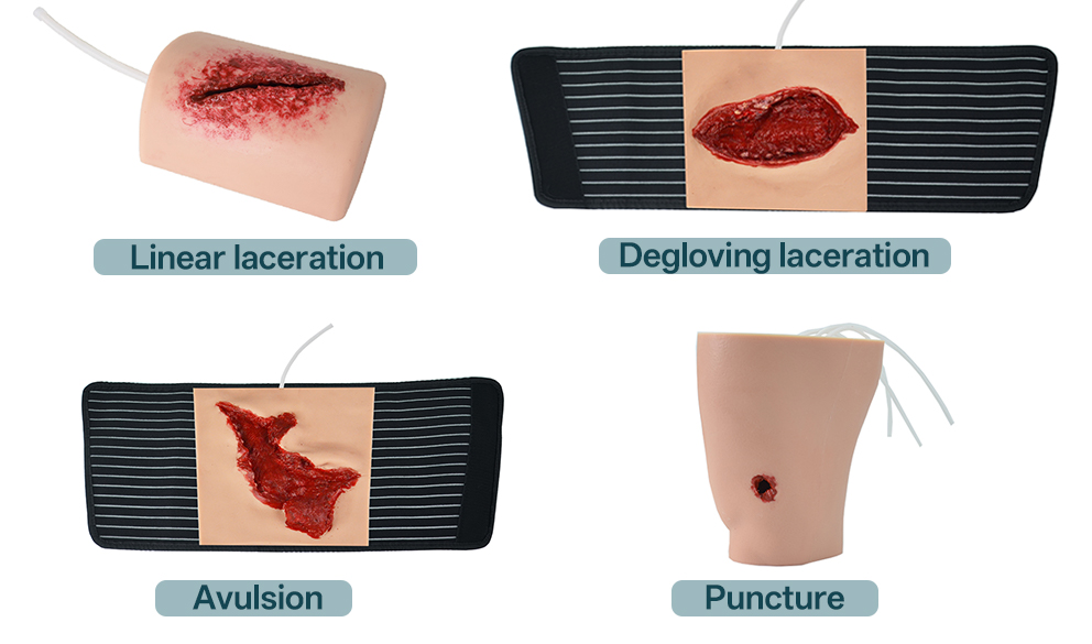
Laceration vs Incision, Avulsion, Abrasion, Puncture, and Contusion
These words describe different injury mechanisms. They can occur together, so a real wound may need more than one term.
| Wound type | Main mechanism | Typical appearance | Key assessment concern |
|---|---|---|---|
| Laceration | Traumatic tearing or splitting | Linear, jagged, stellate, flap, or crushed edges | Depth, contamination, and damage to vessels, nerves, tendons, joints, or bone |
| Incision | Deliberate or accidental cut by a sharp edge | Usually straight, sharply divided edges | Depth and which structures cross the wound path |
| Avulsion | Tissue is forcibly pulled from its attachment | Partially attached flap or complete tissue loss | Blood supply, tissue viability, exposed structures, and hemorrhage |
| Abrasion | Friction scrapes the surface | Broad, shallow, raw area with epidermal loss | Embedded dirt, pain, surface area, and deeper injury beneath road rash |
| Puncture | Narrow object penetrates tissue | Small surface opening with a potentially deep tract | Hidden depth, retained material, contamination, and injury below the opening |
| Contusion | Blunt force damages tissue without necessarily opening skin | Bruising, swelling, tenderness, discoloration | Deeper bleeding, fracture, crush injury, and evolving swelling |
Laceration vs incision
An incision is produced by a sharp edge and usually has more regular margins. A surgical incision is planned and made under controlled conditions. A traumatic incision from glass or metal may look neat but can still be contaminated and can divide deep structures.
A laceration emphasizes traumatic tearing. Its edges may be irregular or crushed, although some lacerations are also linear. The practical lesson is that edge shape does not replace exploration and functional assessment.
Avulsion vs laceration
A laceration separates tissue along a tear. An avulsion pulls tissue partly or completely away from its attachment. A flap laceration sits near the boundary between the terms: the tissue remains connected, but its survival depends on the remaining blood supply.
Avulsion injuries deserve special attention when the flap is pale, dusky, cool, or attached by a narrow bridge; when tissue is missing; or when bone, tendon, cartilage, or another deep structure is exposed.
Abrasion vs laceration
An abrasion is primarily a surface-scraping injury. A laceration opens the tissue through a tear or split. Road rash can include both: a wide abrasion across the surface and one or more deeper lacerations containing gravel or other debris.
This distinction matters in training because the longest or most painful-looking area is not always the deepest injury.
Puncture vs laceration
A puncture wound has a small opening relative to the depth of its tract. A laceration usually exposes more of the wound path. The small surface size of a puncture can be misleading: contamination or damage may sit well below the visible opening.
Punctures, bites, and wounds containing devitalized or contaminated tissue also change the assessment of tetanus exposure risk. Vaccination decisions depend on the wound category and the patient’s immunization history, not on wound size alone.
Contusion vs laceration
A contusion is tissue damage from blunt force, usually with intact skin. A contused laceration combines both patterns: blunt force bruises and crushes tissue while splitting the skin. The surrounding zone of injury may be wider than the open wound suggests.
A Deeper Laceration Assessment: Look Beyond Length
Length is easy to measure, but it is rarely the most important feature. A short wound over a tendon, joint, major vessel, eyelid margin, or fingertip can be more consequential than a longer superficial wound elsewhere.
1. Mechanism and energy
Ask what happened, what object was involved, how fast it was moving, and whether there was crushing, shearing, twisting, or penetration. Mechanism helps predict debris, tissue loss, fracture, and the direction of deeper injury.
2. Hemorrhage and perfusion
Active bleeding should be controlled before detailed examination. The tissue beyond the wound should then be checked for color, warmth, capillary refill, and pulses where appropriate. A dusky flap or poorly perfused distal tissue requires urgent review.
3. Nerves, tendons, joints, and bone
Assessment includes sensation and active movement, ideally before local anesthetic obscures the neurological examination. A partial tendon injury may cause pain or weakness without an obvious resting deformity. Deep wounds near joints may penetrate the joint even when the opening looks small.
4. Foreign bodies and contamination
Glass, gravel, wood, metal, clothing, soil, and bite contamination require different levels of suspicion. No imaging method detects every foreign body, so the history, symptoms, exploration, and imaging findings must be considered together.
5. Tissue viability
Healthy-looking length is not the same as viable tissue. Crushed, macerated, pale, or narrowly based tissue may not have reliable perfusion. Debridement decisions should preserve viable structures while removing tissue that cannot recover; this is a clinical judgment, not a cosmetic attempt to make every wound edge straight.
6. Patient and location factors
Diabetes, vascular disease, immune compromise, medications, delayed presentation, and previous wound-healing problems can alter risk. Location also changes the stakes: injuries involving the hand, face, eye, mouth, genital region, major joints, or areas with limited blood supply may need specialty care.
Does Every Laceration Need Stitches?
No. Closure is chosen after assessment, cleaning, removal of foreign material, and evaluation of tissue viability.
- Primary closure brings suitable wound edges together with sutures, staples, adhesive, or strips.
- Secondary intention leaves a wound open to fill and epithelialize when approximation is not appropriate.
- Delayed primary closure allows a contaminated or higher-risk wound to be cleaned and observed before later closure.
A small, superficial, non-gaping laceration may need only an appropriate dressing. A puncture is often not closed at the surface. A contaminated, infected, crushed, bitten, or devitalized wound may need a different plan from a clean linear laceration. Closure method and timing should therefore follow the wound assessment, not the other way around. In training, a laceration wound packing trainer can help learners distinguish a deep, packable cavity from a superficial wound that requires a different response.
When a Traumatic Wound Needs Urgent Evaluation
Seek urgent medical assessment when a wound has any of the following:
- Bleeding that is severe or does not stop with appropriate direct pressure
- Pale, blue, cool, or poorly perfused tissue beyond the injury
- Numbness, weakness, loss of movement, or severe pain with movement
- Visible fat, fascia, muscle, tendon, bone, or another deep structure
- A deep tract, suspected joint penetration, open fracture, or amputation
- Embedded glass, metal, gravel, wood, or another foreign object
- An animal or human bite, heavy contamination, or significant crush mechanism
- A large flap, degloving injury, tissue loss, or signs that a flap is not viable
- Increasing redness, warmth, swelling, drainage, odor, fever, or worsening pain
- Uncertain tetanus vaccination status, especially with a dirty, puncture, crush, or avulsion wound
Sources
- MSD Manual Professional – Skin Lacerations
- Royal Children’s Hospital Melbourne – Clinical Practice Guideline: Lacerations
- CDC – Clinical Guidance for Wound Management to Prevent Tetanus
- NCBI Bookshelf – Wound Assessment
- NCBI Bookshelf – Abrasion
- WHO training material – Wound classification
This article is for education and simulation training and is not a substitute for professional medical assessment or treatment.