A tension pneumothorax is one of the few conditions in emergency medicine that can kill a patient in minutes yet be reversed in seconds with the right action. This guide explains what a tension pneumothorax is, how it differs from a simple (ordinary) pneumothorax, the signs and symptoms that should raise the alarm, and why it demands immediate decompression. It is the clinical companion to our step-by-step guide on needle decompression sites and technique.
Important: This article is educational and written for healthcare students, clinicians, and pre-hospital providers. It is not a guide to treating yourself or others without training. Anyone with sudden, severe breathing difficulty or chest pain after an injury needs emergency medical care immediately — call your local emergency number.
What Is a Pneumothorax?
A pneumothorax is air trapped in the pleural space — the thin gap between the lung and the chest wall. Normally that space holds only a film of fluid and a slight vacuum that keeps the lung expanded against the chest wall. When air leaks into it — through a hole in the lung or a wound in the chest wall — that vacuum is lost and the lung collapses away from the chest wall, partly or completely.
A collapsed portion of lung can no longer take part in breathing, so the patient becomes short of breath and the affected side moves less. How dangerous this is depends entirely on how much air gets in and whether it can escape again — which is exactly what separates a simple pneumothorax from a tension pneumothorax.
Simple Pneumothorax vs Tension Pneumothorax: The Key Difference
People often search for the difference between a “simple” and a “tension” pneumothorax, and it is the single most important distinction to understand because it decides how urgent the situation is.
- Simple (closed) pneumothorax. Air enters the pleural space and the lung collapses to some degree, but the leak does not keep adding pressure. The volume of trapped air stays relatively stable. The patient may be breathless and in pain, but they are usually not in immediate danger of collapse. Many simple pneumothoraces are managed with observation, oxygen, or a chest drain.
- Tension pneumothorax. Here the leak behaves like a one-way valve: air is pushed into the pleural space with every breath but cannot get back out. Pressure therefore builds progressively with each breath. The trapped air not only collapses the lung on that side but eventually pushes the heart and central structures (the mediastinum) toward the opposite side and squeezes the large veins returning blood to the heart. This is what turns a survivable injury into a rapidly fatal one.
In short: a simple pneumothorax is a collapsed lung; a tension pneumothorax is a collapsed lung plus a pressure problem that starves the heart of blood. The first is often urgent; the second is immediately life-threatening.
Open vs Closed Pneumothorax: Another Way to Classify It
Simple versus tension describes the pressure inside the chest. A second, equally important way to classify a pneumothorax is by whether the chest wall has been breached — this is the open versus closed distinction.
- Closed pneumothorax. The chest wall is intact — there is no wound on the outside. Air reaches the pleural space from within: a ruptured air blister (bleb) on the lung surface, or a fractured rib puncturing the lung. A spontaneous, simple pneumothorax is typically a closed one.
- Open pneumothorax. There is an open wound through the chest wall — usually a penetrating injury such as a stab or gunshot — that connects the pleural space directly to the outside air. Air is drawn in and out through the wound with each breath, which is why it is classically called a “sucking chest wound.”
| Closed pneumothorax | Open pneumothorax | |
|---|---|---|
| Chest wall | Intact — no external wound | Open wound through the chest wall |
| Where the air comes from | Internal (lung or airway leak) | Through the wound from outside (and the lung) |
| Typical cause | Ruptured bleb, rib fracture, spontaneous | Penetrating trauma (stab, gunshot) |
| Hallmark | No visible wound | “Sucking” wound, sometimes bubbling air |
| First response | Treat the cause; observe and monitor | Apply a chest seal over the wound; watch for tension |
The two systems overlap rather than compete: a pneumothorax is either open or closed, and either type can progress to a tension pneumothorax if a one-way valve forms and pressure builds. This matters for first aid — the immediate treatment for an open (“sucking”) chest wound is a chest seal applied over the wound, but it must be watched closely, because a fully occlusive seal with no vent can itself trap air and tip a casualty into tension physiology.
How a Tension Pneumothorax Develops: The One-Way Valve
The whole problem comes down to that one-way valve. A tear in the lung surface or a wound through the chest wall can create a flap of tissue that opens when pressure rises (during inspiration or a positive-pressure breath) and seals shut when pressure falls. Air is drawn or pushed in, the flap closes, and none of it leaves.
As the trapped volume grows, the consequences cascade:
- The lung on the affected side collapses completely, removing it from gas exchange.
- Pressure pushes the mediastinum — the heart, great vessels, and windpipe — toward the unaffected side, which begins to compress the other lung too.
- The large veins (vena cava) are kinked and compressed, so blood can no longer return to the heart effectively.
- Cardiac output falls because the heart has less blood to pump — this is obstructive shock, and it is what actually kills the patient, often before oxygen levels alone would.
This is why a tension pneumothorax can deteriorate so fast: it is a combined breathing and circulation emergency.
What Causes a Tension Pneumothorax?
A tension pneumothorax can arise in several settings:
- Chest trauma — penetrating injuries (stab or gunshot wounds) and blunt trauma (rib fractures puncturing the lung) are the classic causes, especially in pre-hospital and battlefield care.
- Positive-pressure ventilation — a patient on a mechanical ventilator, or being bag-mask ventilated, can develop tension physiology quickly because every delivered breath forces more air through the leak.
- During CPR or resuscitation, where chest compressions and ventilation combine.
- As a complication of medical procedures (central line placement, lung biopsy) — a so-called iatrogenic pneumothorax.
- Spontaneously, when an existing simple or spontaneous pneumothorax progresses to tension — less common, but it does happen, including in people with underlying lung disease.
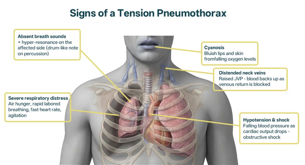
Signs and Symptoms of a Tension Pneumothorax
A tension pneumothorax is, above all, a clinical diagnosis — in a crashing patient you act on the signs at the bedside, not on an X-ray. The picture evolves, so it helps to think in terms of earlier and later signs.
Earlier signs (the patient is struggling but compensating):
- Severe shortness of breath and a feeling of air hunger
- Sharp chest pain on the affected side
- Rapid, labored breathing (tachypnoea) and a fast heart rate (tachycardia)
- Anxiety, restlessness, or agitation from low oxygen
- Reduced or absent breath sounds on the affected side when listening with a stethoscope
- Hyper-resonance (a drum-like note) when tapping the affected side of the chest
Later signs (the patient is decompensating — act immediately):
- Tracheal deviation — the windpipe is pushed away from the affected side (a late, classic, but not always present sign)
- Distended neck veins (raised jugular venous pressure) from blood backing up
- Low blood pressure (hypotension) and signs of shock
- Cyanosis — a bluish tinge to the lips and skin
- Falling oxygen saturation and, ultimately, cardiac arrest if untreated
Waiting for the late signs is dangerous. In a trauma patient with severe breathing distress, absent breath sounds on one side, and shock, you should treat for tension pneumothorax before it reaches tracheal deviation and collapse.
How Is a Tension Pneumothorax Diagnosed?
In an emergency, a true tension pneumothorax should be diagnosed and treated clinically — you do not wait for imaging. Sending an unstable patient for a chest X-ray can cost them their life.
Where time and stability allow, additional tools help confirm the picture:
- Point-of-care ultrasound (POCUS) can rapidly show an absent “lung sliding” sign and is increasingly used in emergency and pre-hospital settings.
- Chest X-ray is appropriate only for a stable patient where the diagnosis is uncertain — it will show the collapsed lung and mediastinal shift, but it is confirmatory, not a prerequisite for treatment in a crashing patient.
The practical rule is simple: in a patient with the clinical signs above and circulatory collapse, treat first.
Why Immediate Decompression Is the Priority
Because the core problem is trapped, rising pressure, the life-saving step is to let that pressure out. Releasing the trapped air converts a tension pneumothorax back into a simple (open) pneumothorax — the lung is still collapsed, but the deadly pressure on the heart and great vessels is relieved and blood can return to the heart again.
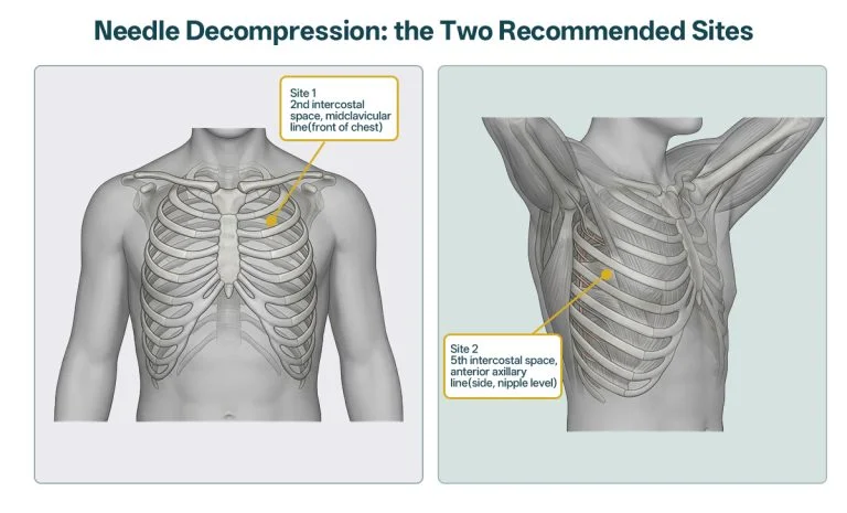
In the field and the resuscitation room this is done first by needle decompression (a needle/catheter inserted into the pleural space to vent the pressure), often followed by finger thoracostomy and a chest drain for definitive management. The technique, the correct insertion sites, and the evidence behind them are covered in detail in our companion guide:
→ Needle Decompression: Sites, Technique, and Step-by-Step Guide
And where no medical kit is available, the same life-saving principle can sometimes be applied with improvised equipment — see our field guide on needle chest decompression with improvised tools.
Frequently Asked Questions (FAQ)
How quickly can a tension pneumothorax become fatal?
There is no fixed clock, but a true tension pneumothorax can progress from breathing distress to circulatory collapse within minutes, particularly in a ventilated patient where every delivered breath adds pressure. That speed is exactly why it’s treated clinically and decompressed on the spot rather than after imaging.
Can a tension pneumothorax happen without an injury?
Yes. While trauma is the classic cause, it can also develop from positive-pressure ventilation, as a complication of procedures such as central-line placement or lung biopsy, or when a spontaneous pneumothorax (sometimes in people with underlying lung disease) progresses to tension. Any of these can create the same one-way-valve effect.
Is a tension pneumothorax the same as a collapsed lung?
Not quite. Every tension pneumothorax involves a collapsed lung, but not every collapsed lung is under tension. The defining extra feature is the progressive pressure that shifts the heart and chokes off venous return — that pressure, not the collapse alone, is what makes it immediately life-threatening.
Can you fully recover after a tension pneumothorax?
When it is recognised and decompressed in time, the outlook is generally good and many people recover fully after the air is drained and the lung re-expands. The danger lies almost entirely in delay — the longer the heart is starved of blood, the worse the outcome.
Why does the trachea move in a tension pneumothorax?
The rising pressure on the affected side physically pushes the central chest structures, including the windpipe, toward the opposite side. Tracheal deviation is a recognised sign, but it appears late and isn’t always present, so its absence never rules out a tension pneumothorax.
Sources
- Advanced Trauma Life Support (ATLS), American College of Surgeons — thoracic trauma and tension pneumothorax
- PHTLS (Prehospital Trauma Life Support) — chest injury management
- StatPearls / NCBI Bookshelf — Tension Pneumothorax
- NHS — Collapsed lung (pneumothorax)
- Resuscitation Council and emergency medicine guidance on chest decompression
This article is for general medical education only. It is not a substitute for accredited clinical training or for professional medical advice. Emergency procedures described here are performed by trained clinicians and pre-hospital providers. If you suspect a serious chest injury, call your local emergency number immediately.
🩺 For Students, EMS, and Training Teams
Recognising and treating a tension pneumothorax is a high-stakes skill that should be drilled long before it’s needed on a real patient. Our Tension Pneumothorax & Needle Decompression Simulator lets students practise identifying landmarks and performing decompression repeatedly, and our Pneumothorax Haemothorax Chest Drain Model With Replaceable Skin Pads supports repeated chest-landmark identification, pneumothorax and haemothorax decompression, and chest-tube insertion practice using replaceable puncture modules.