A difficult dog blood draw is rarely solved by pushing the needle farther or trying more times. Poor jugular filling, unstable restraint, patient movement, excessive syringe suction, partial needle placement, and an unsuitable collection system can all produce the same frustrating result: no blood, interrupted flow, a damaged sample, or a growing hematoma.
This guide is a troubleshooting companion to Dog Jugular Vein Phlebotomy: Positioning, Landmarks, and Step-by-Step Technique. It focuses on recognizing the failure pattern, stopping before harm accumulates, and adapting the plan for small, senior, obese, dehydrated, fearful, or high-bleeding-risk dogs.
Scope: Troubleshooting here is limited to canine jugular blood collection. For peripheral IV catheter placement, medication-route decisions, or feline IV practice, use the separate canine IV catheterization guide, dog SC vs IM guide, or Veterinary Cat IV Practice Kit.
Important: Jugular venipuncture is for trained veterinary personnel and supervised learners. A difficult draw should be escalated to the responsible veterinarian or an experienced team member. Do not attempt to draw blood from a dog at home.
Start With the Failure Pattern
“I cannot get blood” is not a diagnosis. Identify what happened before changing the needle position.
| What you observe | Likely categories to check first | Safer response |
|---|---|---|
| Vein cannot be seen or palpated | Position, occlusion point, coat, skin mobility, poor venous filling | Keep the needle capped; reset the dog, head, thumb, and lighting |
| Blood flashes, then stops | Bevel partly in the lumen, back wall contacted, patient moved, vein collapsed | Reduce suction and reassess alignment; avoid wide blind redirection |
| Syringe is difficult to pull | Excessive vacuum, bevel against vessel wall, small/collapsed vein | Relax suction; stabilize; reconsider syringe or collection system |
| Swelling develops during aspiration | Needle has left or passed through the vein | Stop immediately, release occlusion, withdraw, and apply pressure |
| Separated serum/plasma is pink or the laboratory flags hemolysis | Traumatic collection, strong suction, forcing blood through needle, shaking | Review collection and transfer technique; redraw if the lab requires it |
| Sample clots or has platelet clumps | Slow transfer, delayed mixing, repeated trauma, wrong tube handling | Follow laboratory tube and mixing instructions; recollect when invalid |
The table is a triage aid, not permission to keep probing. When the cause is uncertain, withdraw and reset.
Problem 1: The Jugular Vein Will Not Fill
Before concluding that the dog has a “bad vein,” check the setup.
Recheck the occlusion point
The external jugular lies lateral to the trachea and is occluded near the thoracic inlet. A thumb placed too close to the trachea, too cranially, or without enough contact may not trap venous blood. Release pressure, repalpate the jugular groove, and occlude again while watching for the vessel to rise and soften.
Reset the head and neck
The nose may need to move slightly up, down, or toward the opposite side. Excessive extension is not a universal fix. It can cause discomfort and can pull the skin and soft tissues into an unhelpful position. Aim for comfortable alignment that exposes the groove while preserving normal breathing.
Stabilize mobile skin
Loose skin can move over the vessel and make the landmark appear to shift. Once the vein is identified, use gentle skin tension to stabilize it. Do not mistake a fixed muscle edge or the trachea for a vessel; the jugular should change with occlusion and release.
Improve access to the landmark
Parting or wetting the coat may be enough. Clipping can help with a dense coat, poor visualization, suspected repeated attempts, culture collection, or a practice protocol that requires it. Palpation still matters: a visible line is not proof of a distended vein.
Problem 2: There Is a Flash, but Blood Will Not Continue
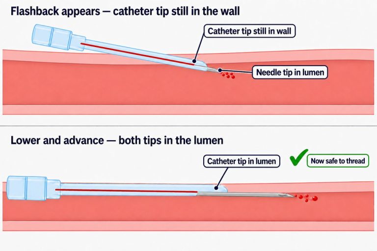
A flash confirms that the needle contacted blood; it does not guarantee that the full bevel remains in the lumen.
First, reduce syringe suction. Strong negative pressure can pull the wall of a small or underfilled vein against the bevel. Confirm that the dog and needle have not moved and that occlusion remains effective.
If a tiny controlled adjustment is part of your supervised protocol, it should follow the vessel’s axis. Avoid sweeping the needle from side to side. Wide redirection creates tissue trauma, makes the final needle position difficult to understand, and raises the risk of hematoma.
When stable flow cannot be restored promptly, release occlusion and suction, withdraw, and apply pressure. A clean reset is safer than prolonged searching beneath the skin.
Problem 3: The Vein Collapses During Aspiration
Vein collapse is more likely when a large evacuated tube is attached to a small vessel, when the patient has poor venous filling, or when a syringe plunger is pulled too aggressively.
Use the least negative pressure that maintains flow. A smaller syringe may give finer control for a small patient, but equipment choice must still allow collection of the required volume and correct tube fill. If an evacuated system repeatedly collapses the vessel, the clinician may choose a syringe or butterfly system that provides more control.
Do not compensate for collapse by repeatedly moving the needle. Pause and determine whether the collection method matches the patient.
Problem 4: A Hematoma Begins to Form
Visible or palpable swelling means blood is entering the surrounding tissue. Stop the draw immediately:
- Release pressure at the thoracic inlet.
- Relax suction on the syringe.
- Withdraw the needle smoothly.
- Apply direct pressure with folded gauze.
- Maintain pressure until bleeding has stopped; do not repeatedly lift the gauze to inspect.
- Reassess the neck and the dog’s breathing before considering another site.
Do not puncture through a forming hematoma or make repeated attempts in the same damaged region. Virginia Tech lists fewer attempts at one site, release of suction and vessel compression before withdrawal, and post-draw compression as core prevention measures.
A rapidly expanding cervical swelling, continued bleeding, respiratory noise, increased breathing effort, pale mucous membranes, weakness, or collapse requires immediate veterinary response.
Preventing Hemolysis and Other Sample Errors
A technically successful stick can still produce an unusable specimen. Hemolysis can alter chemistry results and invalidate parts of a hematology assessment.
Collection errors that damage red cells
- Pulling the syringe plunger too hard or with fluctuating pressure
- Using repeated needle movements that traumatize blood and tissue
- Forcing blood from the syringe through a narrow attached needle
- Expelling blood forcefully against the bottom of a tube
- Shaking rather than gently mixing an additive tube
- Using wet or contaminated equipment
Remove the needle before transferring a syringe sample when the collection system and protocol allow. Let blood run gently down the tube wall, fill the tube to its required level, and mix it according to the receiving laboratory’s instructions.
Underfilled anticoagulant tubes
Underfilling is not a cosmetic problem. Cornell notes that partially filled EDTA tubes can alter cell morphology and indices. Coagulation tubes require the correct blood-to-citrate ratio; a short-filled tube can make the result invalid. Plan the volume before the draw and use a tube size appropriate for the patient and ordered tests.
Clotting and platelet clumps
Have tubes ready, transfer promptly, and gently mix anticoagulant samples without delay. Repeated traumatic attempts can also activate platelets and coagulation. If the sample contains clots, laboratory-confirmed hemolysis, or an incorrect citrate ratio, follow the laboratory’s recollection policy rather than trying to interpret compromised data.
Special Patient: Small and Toy-Breed Dogs
Small dogs have less room for hand placement, smaller circulating blood volume, and veins that may collapse under an aggressive vacuum. The answer is not simply a smaller needle.
- Position the dog where the team can support the whole body and control the front legs without suspending or twisting the neck.
- Consider lateral recumbency when it provides better comfort and control.
- Use equipment that gives controlled aspiration and matches both the vessel and required sample volume.
- Select correctly sized collection tubes so the required additive ratio can still be achieved.
- Ask the laboratory which tests and minimum volumes are truly necessary before collecting.
- Stop early when the vein is traumatized; repeated attempts consume limited sites and increase stress.
Virginia Tech specifically notes that an assistant may need to control the front legs of a small dog during jugular positioning. The restraint should support the patient, not stretch it.
Special Patient: Senior Dogs
Age alone does not prohibit a jugular draw. The limiting factors are often cervical pain, arthritis, reduced range of motion, cardiopulmonary disease, fragile skin, anxiety, or medications that affect hemostasis.
Use a padded, non-slip surface and choose sitting, standing, or lateral positioning based on what the dog can maintain comfortably. Do not force the head to a textbook angle when a smaller adjustment exposes the groove safely. Review medications and recent platelet or coagulation information when relevant.
For a frail senior, a smaller diagnostic plan or a different vein may be preferable to prolonged restraint for an idealized jugular sample. The veterinarian should balance sample quality against the physiologic and behavioral cost of collection.
Special Patient: Obese Dogs, Loose Skin, or a Dense Coat
In these patients, visual landmarks are unreliable and the skin may slide over the vein.
Palpate from the angle of the mandible toward the thoracic inlet, then confirm that the structure changes with occlusion and release. Part or clip the coat as appropriate, and use skin tension after locating the vessel. Repositioning is usually more productive than increasing needle depth based on guesswork.
If the vein cannot be confidently mapped before the needle is uncapped, ask a more experienced operator or choose another method. Blind deep probing in the neck is not acceptable troubleshooting.
Special Patient: Dehydrated, Hypotensive, or Critically Ill Dogs
Poor circulating volume can make peripheral and jugular veins less distended. These patients also have less tolerance for repeated blood loss, stress, and delays in stabilization.
Define the minimum diagnostic volume, prepare every tube in advance, and use the most experienced available operator. Apply gentle, controlled aspiration rather than strong suction. The clinician may choose a different vein, point-of-care testing, ultrasound-guided access, or sampling from an appropriately managed vascular catheter depending on the clinical situation.
Do not assume that the jugular is always the answer to poor peripheral filling. Site choice must fit the resuscitation plan, current vascular access, and airway and bleeding risks.
Special Patient: Fearful, Reactive, or Brachycephalic Dogs
Jugular restraint places hands close to the head and may require neck positioning that a fearful or respiratory-compromised dog cannot tolerate.
Use behavior cues to decide whether the procedure can continue safely. A non-slip surface, side-on handling, minimal restraint, food reinforcement when permitted, and a rehearsed team sequence can reduce escalation. Research on adaptive, collaborative veterinary handling supports reducing unnecessary restraint and tailoring procedures to the dog’s response.
For a brachycephalic dog or any patient with respiratory distress, avoid compressing the airway or forcing the head and neck. A different site, oxygen support, anxiolysis, sedation, or postponement may be safer. Staff safety and patient ventilation take priority over the preferred collection site.
Special Patient: Suspected Coagulopathy
Do not treat this as an ordinary difficult stick. Virginia Tech lists suspected coagulation(blood clotting) disorders as a contraindication to jugular venipuncture because a hematoma or hemorrhage in the neck can compromise the upper airway. Cornell likewise recommends peripheral rather than jugular sampling in patients with severe hemostatic defects.
The veterinarian should select the safest accessible site and collection method. Prepare gauze before puncture, minimize attempts, and maintain pressure for as long as clinically required. A coagulation sample also needs an atraumatic draw and an exact citrate-to-blood ratio; if the tube is short-filled or the sample clots, it should be recollected according to laboratory instructions.
When to Abandon the Jugular Site
Choose another plan when:
- A coagulation disorder or serious bleeding risk is suspected
- Neck trauma, infection, swelling, or an existing jugular device makes the site unsuitable
- The dog cannot tolerate safe head and neck positioning
- Respiratory compromise worsens with restraint
- A hematoma or continued bleeding develops
- The vein cannot be confidently located before puncture
- Controlled attempts have failed and tissue trauma is accumulating
- The available equipment cannot collect the required specimen without collapsing the vein
Escalation is a clinical skill, not a failure. A different operator or site protects the patient and often produces a better sample faster.
Conclusion
A difficult canine jugular draw should trigger diagnosis, not persistence. Identify whether the problem is positioning, occlusion, unstable needle placement, excessive vacuum, poor venous filling, sample transfer, or an unsuitable patient. Reset with the needle capped, use a clear stop rule, and move to another operator, site, or collection plan before tissue trauma and stress accumulate.
For broader site selection and comparison with the cephalic and saphenous veins, return to the canine venipuncture and blood collection guide. For IV access rather than diagnostic sampling, see How to Place a Canine IV Catheter.
Frequently Asked Questions
Can the same jugular vein be used again after a failed blood draw?
Do not immediately repuncture through swelling, bruising, or a forming hematoma. Release occlusion, apply pressure, and let the responsible clinician assess the site. Depending on the tissue, bleeding risk, and urgency of testing, the next attempt may use a fresh area, the opposite jugular, a peripheral vein, or a different collection method.
Should a dog wear a neck bandage after jugular venipuncture?
Routine draws are often managed with direct manual pressure until bleeding stops rather than a take-home neck wrap. Never apply a tight neck bandage yourself: compression or a slipping wrap can interfere with breathing. If the veterinary team places a dressing, follow its removal instructions and contact the clinic promptly for increasing swelling, bleeding, respiratory noise, or breathing difficulty.
Can a blood sample be taken from an existing IV catheter instead?
Sometimes, but the answer depends on the catheter, fluids or drugs running through it, the test ordered, and the clinic or laboratory protocol. Catheter samples can be diluted or contaminated if collection and discard procedures are not followed. The veterinary team should decide whether a catheter sample is valid or whether direct venipuncture is required.
Sources
- Virginia Tech – SOP: Venipuncture in Dogs and Cats
- University of Minnesota – Dog Restraint for Jugular Blood Draw
- Cornell University College of Veterinary Medicine – Comparative Coagulation Sampling Instructions
- Cornell University College of Veterinary Medicine – Hematology Sample Guidance
- Diagnostic and Therapeutic Procedures – Venipuncture Complications and Sample Handling
- Effects of Changing Veterinary Handling Techniques on Canine Behaviour and Physiology
- Comparison of Values Obtained via Jugular Venipuncture and Peripheral IV Catheters in Dogs
This article is for veterinary education and supervised clinical training. It does not replace patient-specific assessment, laboratory instructions, or hands-on instruction by a qualified veterinary professional.
Training Resources
For jugular-specific troubleshooting, practise landmark-based positioning, flashback recognition, needle control, and controlled aspiration with MEDTACEDU’s Canine Jugular Venipuncture & Jugular Phlebotomy Practice Model before working with live patients. For peripheral venipuncture and catheter skills, see the Dog IV Practice Kit and Canine IV Training Model. Explore all Veterinary IV & Practice Models.