Most failed feline blood draws are not caused by poor needle technique. They are caused by a decision made before the needle came out of the packet: the wrong vein was chosen for the volume required, for the cat in front of you, or for what that cat is likely to need next.
Choose well and a single stick produces a usable sample. Choose badly and the same cat is restrained twice, the sample clots in the syringe, or the vein that was needed for an IV catheter an hour later has already been used.
This guide covers how the site is chosen. Technique for each site, restraint, and the specific differences between cats and dogs are covered in separate guides.
Quick Answer: Which Vein Should You Use?
Work backward from the sample. Decide how much blood the tests require, then judge what handling this cat will tolerate, then check whether a limb vein needs to stay available for intravenous access. The site follows from those three answers.
The three common feline venipuncture sites are the jugular, medial saphenous, and cephalic veins. The femoral vein is an occasional fallback rather than a routine first choice.
| Site | Typical yield | Best suited to | Main limitation |
|---|---|---|---|
| Jugular | Largest volume, fastest flow | Full hematology and biochemistry panels, multiple tubes, ill patients needing a broad workup | Requires tolerance of neck handling; higher hematoma risk if hemostasis is poor |
| Medial saphenous | Small to moderate | Cats that will not accept neck handling; semilateral positioning; preserving forelimb veins | Small vessel; slow collection invites clotting in the syringe |
| Cephalic | Roughly 1–3 mL | Small single-tube samples | Usually the preferred vein for an IV catheter, so using it may cost you later access |
| Femoral | Small to moderate | Occasional use when other sites have failed | Harder to compress afterwards; greater hematoma risk |
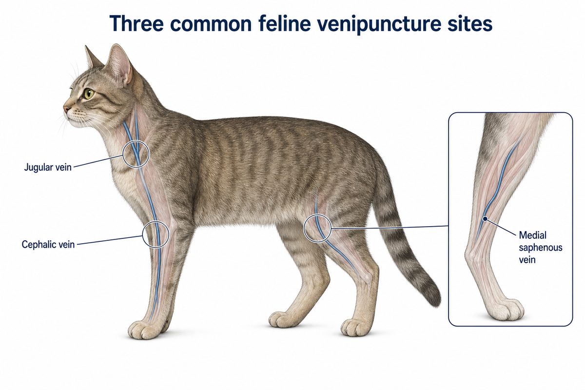
Original educational illustration showing the three feline sites in this table. The hind limb inset is the medial saphenous vein on the inner surface of the leg, not the lateral saphenous vein used routinely in dogs.
One point deserves emphasis before anything else, because getting it wrong marks a handler as inexperienced with cats: the feline hindlimb site is the medial saphenous vein, on the inner surface of the leg. In dogs the routine hindlimb site is the lateral saphenous vein, on the outer surface. They are different vessels approached from opposite sides, and technique transferred directly from canine lateral saphenous collection will not find the feline vein.
Start With the Volume, Not the Vein
The most common planning error is choosing a vein first and then discovering it cannot supply what the laboratory needs.
Begin by adding up the requirement. A hematology tube, a biochemistry tube, and an additional test such as total T4 or a retrovirus screen each need their own fill volume, and tubes have minimum fill requirements that must be respected for the results to be valid. Underfilled anticoagulant tubes produce a dilution artifact; overfilled ones clot. Ask the laboratory what it needs before drawing, not afterwards.
Then check the ceiling. Total blood volume in a cat is usually quoted at roughly 55–70 mL/kg, and the figure varies between references, so use the one your practice protocol specifies. A widely used safety limit for a healthy patient is no more than about 10% of total blood volume in a single collection, which is often expressed clinically as up to about 1% of body weight. For a 5 kg cat that lands in the region of 5 mL. Laboratory-animal guidance commonly pairs a 10% single draw with a recovery interval of about two weeks before the next one.
These are ceilings for healthy patients, and they fall for anemic, dehydrated, unstable, very small, pediatric, and geriatric cats. The volume that is appropriate for a particular patient is a clinical decision made under veterinary direction and according to your practice’s protocol.
Once the number is fixed, the site choice narrows on its own. A full workup on a 4 kg cat points toward the jugular. A single small tube does not.
Jugular Blood Draw in Cats
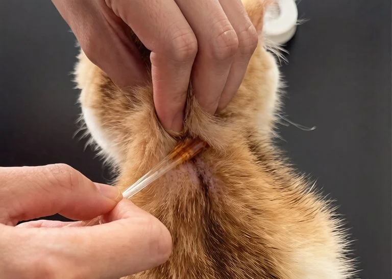
The jugular is the site of choice when volume or speed matters. It is a large vessel, it fills tubes quickly, and a shorter time with the needle in the vein means less opportunity for the sample to be damaged or for the cat to become distressed mid-procedure.
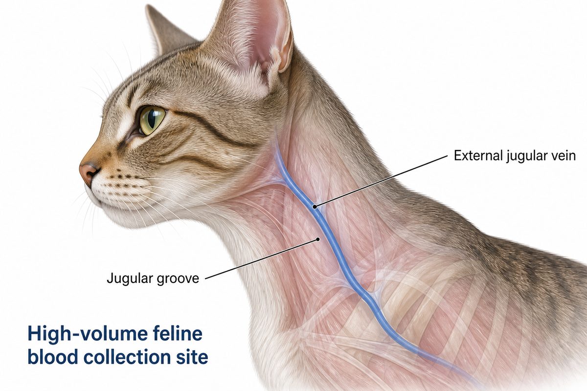
Original educational illustration of the feline jugular groove and external jugular vein. Orientation only; it does not depict puncture technique.
The vein runs in the jugular groove on either side of the neck. A common approach is to begin in the caudal third and, if an attempt fails, move progressively cranially rather than repeatedly puncturing the same spot.
Its limits are practical rather than technical. Neck handling is the part many cats tolerate least, and current feline-friendly guidance favors minimal handling over firm restraint, since forceful restraint measurably increases fear responses and makes the next visit harder. The jugular is also a poor choice where there is a coagulopathy, thrombocytopenia, or local disease, and it demands proper post-collection pressure: a hematoma in the neck of a cat is both more likely and more consequential than one on a limb.
The Medial Saphenous Vein in Cats
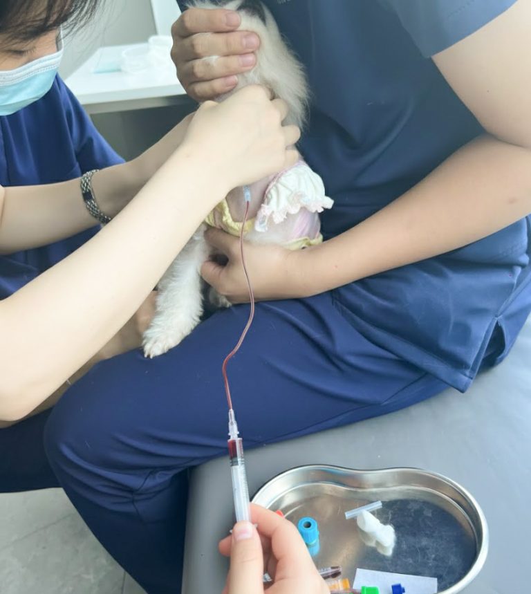
The medial saphenous runs on the inner surface of the hindlimb—the routine cat blood draw location on the back leg—and is, for many practitioners, the most useful feline site after the jugular. It is reached with the cat in lateral or semilateral position, with an assistant lifting the upper hindlimb and applying gentle occlusion proximally, and it avoids neck handling entirely. For a cat that objects to jugular restraint but settles on its side, it is frequently the difference between a sample and no sample.
Its weakness is size. The vessel is small, and collections beyond a couple of milliliters can be slow or simply unobtainable. Slow collection is the real problem: blood that trickles into a syringe over a prolonged period may begin to clot before it reaches the tube, and a clotted sample is a wasted stick. A butterfly set gives finer control and keeps the needle steady while the syringe is handled, and needle gauge should be matched to the vessel rather than to habit — too large a needle collapses a small vein, while excessive suction on any gauge causes hemolysis.
If the requirement is small, the cat will not accept neck handling, and the forelimb veins must be preserved, this is usually the right site.
Cephalic Blood Draw in Cats
The cephalic vein on the dorsal forelimb is the most familiar vessel to anyone trained on dogs, and it is well suited to small samples in the region of 1–3 mL.
The reason to think twice is not technical. In most practices the cephalic vein is also the first choice for an intravenous catheter. If this cat may be admitted, sedated, given fluids, or anesthetized later in the same visit, spending the cephalic vein on a blood sample can mean the person placing the catheter inherits a bruised, punctured vessel and has to move to the other leg or to a less convenient site. Deciding whether the cat is likely to need intravenous access before drawing from the cephalic is a small habit that prevents a recurring problem.
Repeated attempts also cost more here than elsewhere, because the vessel rolls readily in cats and a series of punctures reduces its usability quickly.
The Femoral Vein
The femoral vein is used in some practices when other sites have failed or when a cat’s conformation makes limb access difficult. It is best treated as a site for experienced hands. It is deeper, it is harder to compress effectively afterwards, and hematoma risk is correspondingly higher. It should not be a routine first choice, and it should not be attempted casually because two other sites did not work.
Knowing When to Change Site Instead of Trying Again
Persistence at a single site is one of the most reliable ways to turn a routine visit into a difficult one. A workable discipline looks like this:
- After two unsuccessful attempts at the same site, change something real — a different vein, a different handler, or a different day. A third attempt in the same place rarely succeeds and reliably worsens the cat’s tolerance.
- Stop if a hematoma forms. Apply pressure and move to another site rather than working through a swelling that has already distorted the anatomy.
- Watch the cat rather than only the vein. Escalating fear responses are a reason to pause, not a reason to hold tighter. Rescheduling with a pre-visit plan often costs less time overall than forcing a sample today and inheriting an unhandleable patient at every future visit.
- Where a cat cannot be sampled safely, discuss options with the attending veterinarian rather than escalating restraint.
Protecting the Sample After You Have It
A correctly chosen site can still produce an unusable sample in the last thirty seconds.
Transfer blood into tubes promptly, especially after a slow collection from a small vein, and respect fill lines. Remove the needle before transferring into a tube, or use a transfer device, rather than forcing blood through a fine needle under pressure. Invert anticoagulant tubes gently and fully instead of shaking them. Fill tubes in the order your laboratory specifies. Then get the sample to the analyzer or the courier without unnecessary delay.
Hemolysis and clotting account for a large share of rejected feline samples, and both are usually procedural rather than unavoidable.
Conclusion
Site selection in cats is a sequence, not a preference. Establish the volume the tests require, judge what handling this cat will genuinely accept, and check whether a limb vein must stay clean for intravenous access. Those three answers point to one site more often than not — jugular for volume, medial saphenous when neck handling is refused, cephalic only for small samples and only when catheter access is not in the plan.
Getting that decision right is what separates a single clean stick from a repeated, escalating attempt on a cat that will remember it.
Frequently Asked Questions
Can blood be drawn from a cat’s tail vein? Ventral tail venipuncture is used in some species and occasionally described in cats, but it is not a routine feline collection site in general practice. Yield is limited, positioning is awkward, and most cats object strongly to tail handling. In practice the jugular, medial saphenous, and cephalic veins cover the requirement.
How much blood can safely be taken from a very small or elderly cat? The general ceiling of roughly 10% of blood volume applies to healthy patients. Kittens, very small adults, and cats that are anemic, dehydrated, or systemically unwell need a lower limit, and in those patients it is worth asking the laboratory in advance whether tests can be run on reduced volumes, or prioritizing which tests matter most, rather than collecting the maximum and hoping.
Why does a cat’s blood sample keep clotting in the syringe? Almost always because the collection took too long. Small feline veins deliver blood slowly, and blood begins to clot while it is still in the syringe. Shorten the interval between draw and transfer, use a butterfly set for better control, match needle gauge to the vessel, and move to a higher-flow site such as the jugular when the volume required makes a slow peripheral collection unrealistic.
Sources
- American Association of Feline Practitioners / International Society of Feline Medicine — Cat Friendly Veterinary Interaction Guidelines: Approach and Handling Techniques (catvets.com)
- Merck Veterinary Manual — Collection and Submission of Laboratory Samples from Animals (merckvetmanual.com)
- Veterinary Practice News — Challenges in feline blood collection (veterinarypracticenews.com)
- dvm360 — Tips for technicians to improve phlebotomy skills (dvm360.com)
Disclaimer: This article is for professional education only and is not a substitute for veterinary advice, diagnosis, or treatment. Blood collection volumes, site selection, and handling decisions for an individual patient must follow your practice’s protocols and the direction of the attending veterinarian. Last updated: August 2026.
Training Resources
Site selection is a judgment built on top of a reliable hand sequence, and that sequence is best rehearsed away from a live patient.
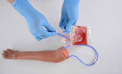
The Veterinary Cat IV Practice Kit is a feline peripheral-vein leg model for practicing vein puncture, blood draw with realistic flashback, catheter placement, and fluid-infusion setup. It represents a limb vein, so it supports peripheral collection and catheter practice; it is not a jugular model and does not simulate neck collection.
For the canine equivalent of this decision, see Canine Venipuncture: Best Practices for Blood Collection.
Coming to cats from dogs? Cat vs Dog Venipuncture: What Actually Changes covers the four defaults that flip, starting with the hind limb site.