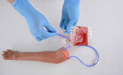
If you are learning to inject filler, the complication you probably worry about most is a vascular occlusion: filler entering an artery and blocking blood flow to the skin, or in rare cases, to the eye. It is the main reason new injectors feel nervous before their first patient, and it is why every aesthetic course teaches the facial danger zones, the areas where an important artery runs close to a common injection site.
The hard part is that most of us learn these arteries from flat diagrams. A diagram shows where an artery usually runs, but not how deep it sits, which bony landmark you can feel to find it, or how far it can stray from the textbook path in a real face. That is exactly what matters at the injection site.
This guide goes zone by zone. For each one it names the arteries, explains where they run and how deep they usually sit, and shows why that zone carries the risk it does. It is written for medical, nursing, and dental students and for new injectors who want the anatomy clear before they treat a patient.
Why Filler in an Artery Can Affect Vision
Every artery in the face comes from the two common carotid arteries, which split into the external and internal carotid arteries. Most of the face is supplied by the external carotid system. The eye is supplied by the ophthalmic artery, which is the first branch of the internal carotid artery.
The two systems are not sealed off from each other. Branches of the ophthalmic artery, such as the supratrochlear, supraorbital, and dorsal nasal arteries, leave the orbit and supply the forehead and upper nose. There they connect with branches of the facial artery, so the ophthalmic artery acts as the main bridge between the two systems.
That bridge explains the most widely accepted mechanism of filler-related blindness. If filler enters one of these arteries, it can travel backward against normal blood flow into the ophthalmic artery, then forward into the arteries that supply the retina. Researchers have reproduced this in the lab. In a 2019 study published in Aesthetic Surgery Journal, Cho and colleagues injected hyaluronic acid filler into the supratrochlear artery of fresh cadaver heads, and it reached the ophthalmic artery in three of six heads.
It also explains why the zones are not equally risky. A 2024 review of vision loss after filler by Doyon and colleagues, published in Aesthetic Surgery Journal, collected 365 cases reported between 2018 and 2023. In those cases, the most common injection sites were the nose (40.6%), the forehead (27.7%), and the glabella (19.0%), all areas supplied at least partly by ophthalmic branches. Blindness after filler is rare, but this pattern shows where anatomy knowledge counts most.
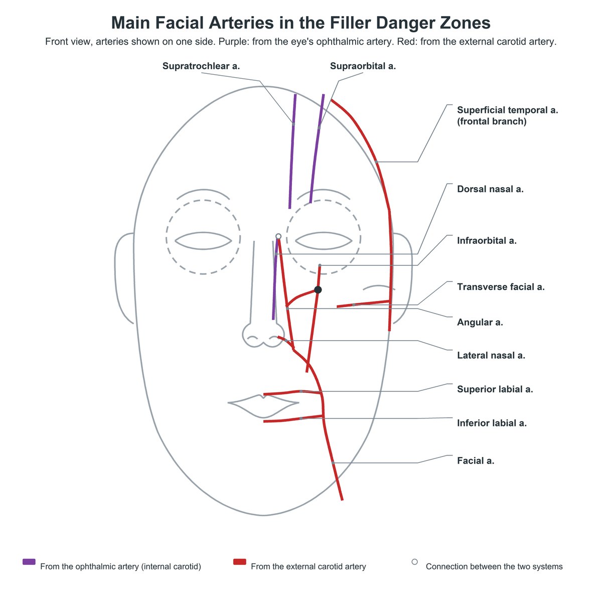
Figure 1. Main facial arteries in the filler danger zones. Purple: branches of the ophthalmic artery. Red: branches of the external carotid artery.
Glabella and Forehead: Supratrochlear and Supraorbital Arteries
The glabella is the area between the eyebrows. Two arteries from the ophthalmic system emerge from the upper rim of the orbit here:
- Supratrochlear artery: runs upward close to the midline. In a 2023 Doppler ultrasound study of 74 half-faces by Shen and colleagues, most supratrochlear arteries ran 1.0 to 2.0 cm from the midline.
- Supraorbital artery: runs upward further out, mainly 2.0 to 4.0 cm from the midline in the same study. It leaves the orbit through the supraorbital foramen.
Both arteries become superficial quickly. In the Cho cadaver study above, the superficial branch of the supratrochlear artery sat only about 1.5 mm below the skin surface. Because they are direct branches of the ophthalmic artery, they offer the shortest route from the skin to the eye, which is why the glabella has ranked among the three highest-risk sites in every published case review.
Temple: Superficial Temporal Artery
The temple is a common site for volume restoration, and the main artery here is the superficial temporal artery, a terminal branch of the external carotid. It runs up the side of the head, and its frontal branch crosses the temple toward the forehead.
In a 2022 cadaver study of the anterior temple by Park and colleagues, a smaller branch called the zygomatico-orbital artery came off the frontal branch in 85.2% of cases and ran along a line from the tragus to the brow. The same authors describe inadvertent injection into the superficial temporal artery as the most common vascular complication of temple filler.
Nose: Dorsal Nasal, Lateral Nasal, Columellar, and Angular Arteries
The nose is the highest-risk site in the two most recent case reviews, largely because its arteries come from both the internal and the external carotid systems and join into a ring around the nose.
- Dorsal nasal artery: a branch of the ophthalmic artery that runs down the bridge of the nose. Over the nasal bone it lies superficial to the muscle layer. Lower down, over the cartilage, it runs deeper.
- Lateral nasal artery: part of the facial artery supply to the side of the nose. Its anterior branch carries most of the blood flow to the nasal tip.
- Columellar artery: supplies the columella, the strip of tissue between the nostrils. It usually comes from the superior labial artery.
- Angular artery: a branch of the facial artery that runs up beside the nose toward the inner corner of the eye. Its course varies widely between people.
A 2012 study by Saban and colleagues, combining Doppler ultrasound with cadaver dissection, showed these vessels form one connected network linking the external and internal carotid arteries, so filler in any of these branches can, in principle, reach the ophthalmic system.
Lips and Perioral Region: Labial Arteries and the Facial Artery
The facial artery runs upward from the lower jaw toward the nose, passing near the corner of the mouth. Around the mouth it gives off two branches:
- Superior labial artery: supplies the upper lip. In a 2015 cadaver study of 60 half-faces by Lee and colleagues, it arose about 12 mm lateral to the corner of the mouth and ran along the vermilion border toward the midline.
- Inferior labial artery: supplies the lower lip, arising near or below the corner of the mouth.
A 2020 ultrasound study of 41 volunteers by Cotofana and colleagues, published in Aesthetic Surgery Journal, found both labial arteries most often in the submucosal plane (58.5%), behind the muscle on the inner side of the lip, and usually within the red part of the lip.
Infraorbital Region and Midface: Infraorbital and Angular Arteries
The infraorbital artery exits the infraorbital foramen, below the lower rim of the orbit, along with the infraorbital nerve. It divides into branches that run toward the lower eyelid, the side of the nose, and the upper lip. In a 2018 cadaver study by Kim and colleagues, the nasal and labial branches were present in every specimen, while the palpebral branch was present in 34.7%.
These branches connect with the supratrochlear, dorsal nasal, and angular arteries. In a 2019 fresh cadaver study, Hufschmidt and colleagues identified two danger zones in this area: the mid-cheek when filler is placed too superficially, and the tear trough and infraorbital hollow when filler is placed deep on the bone. The angular artery runs along the inner edge of this region and links the facial artery to the ophthalmic branches around the eye.
Lateral Cheek and Outer Eye: Transverse Facial and Lateral Palpebral Arteries
The lateral face is usually considered lower risk, but it is not risk-free.
- Transverse facial artery: runs forward across the side of the face below the zygomatic arch. In a 2019 CT angiography study of 200 sides by Koziej and colleagues, it was present in 96% of cases and came from the superficial temporal artery in 91.7%. In some people, a large transverse facial artery continues toward the nose and joins the angular artery, which may explain some cases of blindness after injections in the lateral face.
- Lateral palpebral artery: supplies the outer part of the eyelids. The skin of the outer upper and lower lids is fed by branches of both the superficial temporal artery and the lacrimal artery, which is a branch of the ophthalmic artery, so this small area connects to both carotid systems. Some training models label this vessel “lateral canthal artery”, but lateral palpebral artery is the standard anatomical name.
Facial Danger Zones at a Glance
| Zone | Main arteries | Why it matters |
|---|---|---|
| Glabella and forehead | Supratrochlear, supraorbital | Direct branches of the ophthalmic artery; very superficial |
| Temple | Superficial temporal (frontal branch) | Common site of inadvertent arterial injection |
| Nose | Dorsal nasal, lateral nasal, columellar, angular | Joins the internal and external carotid systems |
| Lips and perioral | Facial, superior labial, inferior labial | Labial arteries run inside the lip |
| Infraorbital and midface | Infraorbital, angular | Branches connect with arteries around the eye |
| Lateral cheek and outer eye | Transverse facial, lateral palpebral | Can link to the angular artery or the ophthalmic system |
How to Learn the Danger Zones Before Your First Patient
Knowing the names is not the same as being able to find the arteries. A few habits close that gap:
- Tie every artery to a landmark you can feel. The orbital rim, the supraorbital notch, the infraorbital foramen, the corner of the mouth, the zygomatic arch, and the tragus are fixed points. Learn where each artery runs relative to them, not relative to a picture.
- Learn the depth, not just the line. The supratrochlear artery becomes superficial within a few millimeters of the skin, while the labial arteries usually run on the inner side of the lip muscle. The layer matters as much as the path.
- Expect variation. The courses in this guide are the usual ones. Several of these arteries are missing or take a different path in a meaningful share of people, so treat the textbook path as a starting point.
- Trace the arteries in three dimensions. Following each vessel from the carotid artery up to its facial branches on a three-dimensional anatomical model shows how the external carotid branches meet the ophthalmic system, which a flat diagram cannot.
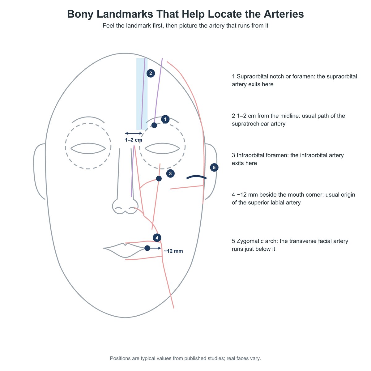
Figure 2. Bony landmarks that help locate the facial arteries. Positions are typical values; real faces vary.
Conclusion
Facial danger zones are not arbitrary lines. Each one marks a place where an important artery runs close to a common injection site, and the most dangerous zones are those whose arteries connect to the ophthalmic artery. That is why the nose, forehead, and glabella lead the case reviews. You do not need to memorize every study figure. Know which arteries sit in each zone, the landmarks that locate them, the layer they usually run in, and how much they can vary, and learn it hands-on before your first patient rather than during it.
Frequently Asked Questions
Is the nasolabial fold a facial danger zone?
Yes. The nasolabial fold is listed among the classic facial danger zones and accounted for 13.3% of cases in the 2015 review of filler-related vision loss and 14.6% in the 2019 update. Both the facial artery and branches of the infraorbital artery supply the area around the fold, and anatomists study their relationship specifically because of nasolabial fold augmentation.
Do facial veins matter, or only arteries?
Veins matter too, though the risks differ. In the temple, the middle temporal vein runs between the layers of the deep temporal fascia. A 2020 systematic review by Kapoor and colleagues links accidental injection into this vein with non-thrombotic pulmonary embolism, where filler travels through the veins to the lungs. Arteries remain the main concern for skin damage and vision loss.
Why do sources give different positions and depths for the same artery?
Facial arteries vary a lot between people, and studies measure them in different ways. Lee and colleagues (2015) found no superior labial artery in 6.7% of half-faces, and Lu and colleagues (2022) found the dorsal nasal artery in only 58% of specimens. Depth also depends on method: the Lee cadaver study placed the superior labial artery about 3 mm deep, while the Cotofana ultrasound study of living volunteers measured about 5.6 mm. Students should learn the usual course of each artery and expect variation in real patients.
Sources
- Doyon VC, Liu C, Fitzgerald R, Humphrey S, Jones D, Carruthers JDA, Beleznay K. Update on blindness from filler: review of prognostic factors, management approaches, and a century of published cases. Aesthet Surg J. 2024;44(10):1091-1104. https://doi.org/10.1093/asj/sjae091
- Beleznay K, Carruthers JD, Humphrey S, Jones D. Avoiding and treating blindness from fillers: a review of the world literature. Dermatol Surg. 2015;41(10):1097-1117. https://doi.org/10.1097/DSS.0000000000000486
- Beleznay K, Carruthers JDA, Humphrey S, Carruthers A, Jones D. Update on avoiding and treating blindness from fillers: a recent review of the world literature. Aesthet Surg J. 2019;39(6):662-674. https://doi.org/10.1093/asj/sjz053
- Scheuer JF 3rd, Sieber DA, Pezeshk RA, Campbell CF, Gassman AA, Rohrich RJ. Anatomy of the facial danger zones: maximizing safety during soft-tissue filler injections. Plast Reconstr Surg. 2017;139(1):50e-58e. https://doi.org/10.1097/PRS.0000000000002913
- Cho KH, Dalla Pozza E, Toth G, Bassiri Gharb B, Zins JE. Pathophysiology study of filler-induced blindness. Aesthet Surg J. 2019;39(1):96-106. https://doi.org/10.1093/asj/sjy141
- von Arx T, Tamura K, Yukiya O, Lozanoff S. The face – a vascular perspective. A literature review. Swiss Dent J. 2018;128(5):382-392. https://doi.org/10.61872/sdj-2018-05-405
- Bird B, Stawicki SP. Anatomy, Head and Neck, Ophthalmic Arteries. In: StatPearls. StatPearls Publishing; updated August 8, 2023. https://www.ncbi.nlm.nih.gov/books/NBK482317/
- Shen WW, Du JN, Ma JX, Xia YC, Cui LG. Evaluation of supratrochlear, supraorbital and angular artery course variations and depth by Doppler ultrasound. Aesthetic Plast Surg. 2023;47(2):791-798. https://doi.org/10.1007/s00266-022-03188-9
- Park HJ, Lee JH, Jung W. The superficial temporal artery and zygomatico-orbital artery: superficial arterial distribution of the anterior temple area. Biomed Res Int. 2022;2022:3790546. https://doi.org/10.1155/2022/3790546
- Choi DY, Bae JH, Youn KH, et al. Topography of the dorsal nasal artery and its clinical implications for augmentation of the dorsum of the nose. J Cosmet Dermatol. 2018;17(4):637-642. https://doi.org/10.1111/jocd.12720
- Lu Y, Hong WJ, Luo CE, Zhan WF, Luo SK. Vasculature of the nasal cartilage region related to filler injection. Aesthetic Plast Surg. 2022;46(5):2461-2468. https://doi.org/10.1007/s00266-022-02942-3
- Saban Y, Andretto Amodeo C, Bouaziz D, Polselli R. Nasal arterial vasculature: medical and surgical applications. Arch Facial Plast Surg. 2012;14(6):429-436. https://doi.org/10.1001/archfacial.2012.202
- Pinar YA, Bilge O, Govsa F. Anatomic study of the blood supply of perioral region. Clin Anat. 2005;18(5):330-339. https://doi.org/10.1002/ca.20108
- Lee SH, Gil YC, Choi YJ, Tansatit T, Kim HJ, Hu KS. Topographic anatomy of the superior labial artery for dermal filler injection. Plast Reconstr Surg. 2015;135(2):445-450. https://doi.org/10.1097/PRS.0000000000000858
- Cotofana S, Alfertshofer M, Schenck TL, et al. Anatomy of the superior and inferior labial arteries revised: an ultrasound investigation and implication for lip volumization. Aesthet Surg J. 2020;40(12):1327-1335. https://doi.org/10.1093/asj/sjaa137
- Kim HS, Lee KL, Gil YC, Hu KS, Tansatit T, Kim HJ. Topographic anatomy of the infraorbital artery and its clinical implications for nasolabial fold augmentation. Plast Reconstr Surg. 2018;142(3):273e-280e. https://doi.org/10.1097/PRS.0000000000004704
- Hufschmidt K, Bronsard N, Foissac R, et al. The infraorbital artery: clinical relevance in esthetic medicine and identification of danger zones of the midface. J Plast Reconstr Aesthet Surg. 2019;72(1):131-136. https://doi.org/10.1016/j.bjps.2018.09.010
- Kim YS, Choi DY, Gil YC, Hu KS, Tansatit T, Kim HJ. The anatomical origin and course of the angular artery regarding its clinical implications. Dermatol Surg. 2014;40(10):1070-1076. https://doi.org/10.1097/01.DSS.0000452661.61916.b5
- Koziej M, Polak J, Wnuk J, et al. The transverse facial artery anatomy: implications for plastic surgery procedures. PLoS One. 2019;14(2):e0211974. https://doi.org/10.1371/journal.pone.0211974
- Toure G, Nguyen TM, Vlavonou S, Ndiaye MM. Transverse facial artery: its role in blindness after cosmetic filler and botulinum toxin injections. J Plast Reconstr Aesthet Surg. 2021;74(8):1862-1869. https://doi.org/10.1016/j.bjps.2020.12.042
- Nguyen JD, Duong H. Anatomy, Head and Neck, Lateral Nasal Artery. In: StatPearls. StatPearls Publishing; updated November 14, 2022. https://www.ncbi.nlm.nih.gov/books/NBK546681/
- Karti O, Saatci I, Saatci AO. Vascular supply of the eye: clinical anatomy. Med Hypothesis Discov Innov Ophthalmol. 2024;13(4):176-189. https://doi.org/10.51329/mehdiophthal1509
- Kapoor KM, Bertossi D, Li CQ, Saputra DI, Heydenrych I, Yavuzer R. A systematic literature review of the middle temporal vein anatomy: ‘venous danger zone’ in temporal fossa for filler injections. Aesthetic Plast Surg. 2020;44(5):1803-1810. https://doi.org/10.1007/s00266-020-01791-2
This article is for education and training purposes only. It describes facial vascular anatomy for teaching and is not a guide to injection technique or to managing complications. Filler injections should be performed only by trained, licensed practitioners.
Training Resources
MEDTACEDU offers two facial injection models that show the arteries in this guide. Students and new injectors can use them to trace each artery from the carotid system to its facial branches and locate every danger zone before treating a real patient, and instructors can use them to teach the same anatomy to a group.
- The Facial Injection Training Head with Muscles, Arteries & Nerves displays the facial muscles, main arteries, and nerves on one side of a silicone face, including the supratrochlear, supraorbital, dorsal nasal, angular, lateral nasal, labial, infraorbital, transverse facial, and superficial temporal arteries, plus the common and internal carotid arteries. It suits courses that teach muscles and vessels together.
- The Clear Facial Injection Model with Vessels shows the arteries, veins, and nerves through a thin, replaceable clear skin and adds the columellar artery. It is the lower-cost option for teaching vascular anatomy to a group.