During a pelvic exam, a clinician may notice that the cervix looks red, swollen, bumpy, enlarged, or different from expected. These changes can be worrying, but many have a benign cause.
It also helps to separate two parts of the visit:
- The speculum exam lets the clinician see the cervix.
- A Pap test collects cervical cells for a laboratory to examine.
A Pap test does not diagnose a visible lump, ulcer, or polyp. In the same way, looking at the cervix cannot confirm cancer or tell its stage. An unusual finding may lead to an HPV test, infection testing, colposcopy, or biopsy.
This guide explains seven cervical appearances that may be seen during a speculum exam and what usually happens next.
What Does a Healthy Cervix Usually Look Like?
The cervix is the lower, narrow part of the uterus. It opens into the vagina. During a speculum exam, it often looks smooth and pink, with a small opening in the center.
Normal appearance varies. Pregnancy, childbirth, menopause, hormones, and the timing of the menstrual cycle can all change the cervix. For example, the central opening may look round in someone who has not given birth vaginally and more slit-like after vaginal birth.
Color alone is not enough to label a cervix normal or abnormal. The clinician also considers symptoms, bleeding, discharge, tenderness, screening history, pregnancy status, and whether a change is new.
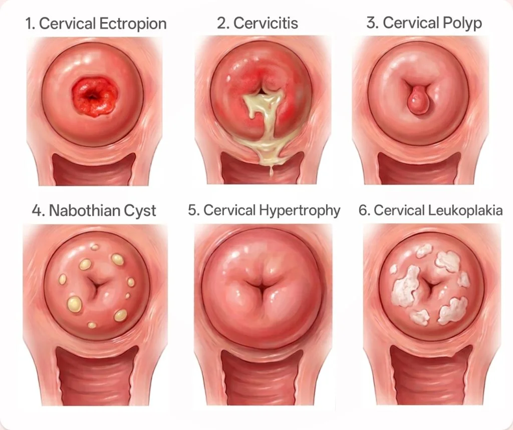
1. Cervical Ectropion: A Red Area Around the Opening
Cervical ectropion, sometimes called cervical ectopy, happens when delicate glandular cells from inside the cervical canal are present on the outer surface.
It often appears as a smooth, bright-red area around the cervical opening. Because these cells are fragile, they may bleed after sex or when touched during an exam. Some people also notice more vaginal discharge.
Ectropion is common during pregnancy, adolescence, and use of estrogen-containing contraception. It is usually a normal, benign change rather than a precancerous lesion.
Treatment is not always needed. If bleeding or discharge is troublesome, the clinician first checks for other causes, including infection and abnormal cells, before discussing treatment.
2. Cervicitis: A Red, Swollen, or Easily Bleeding Cervix
Cervicitis means inflammation of the cervix. The cervix may look red or swollen, bleed easily when touched, or have mucus or pus coming from the opening.
Possible symptoms include:
- unusual vaginal discharge;
- bleeding between periods or after sex;
- pain during sex;
- pelvic discomfort;
- burning or pain with urination.
Chlamydia and gonorrhea are common infectious causes. Trichomoniasis, genital herpes, and Mycoplasma genitalium may also be involved. Sometimes no infection is found. Irritation, changes in the vaginal environment, or another condition can produce a similar appearance.
Appearance alone cannot identify the cause. Testing guides treatment, and sexual partners may also need evaluation when a sexually transmitted infection is found.
3. Cervical Polyp: A Small Growth From the Cervix
A cervical polyp is a soft growth that forms on the cervix or inside the cervical canal. It may look like a smooth red, pink, or purple finger-shaped projection.
Most cervical polyps are benign. Some cause no symptoms and are found during a routine exam. Others cause spotting between periods, bleeding after sex, or increased discharge.
A clinician may recommend removing a polyp if it causes symptoms, looks unusual, or needs laboratory examination. Any bleeding after menopause should be assessed even when a polyp seems to be the likely source.
4. Nabothian Cyst: A Smooth White or Yellow Bump
Nabothian cysts form when mucus becomes trapped under the surface of the cervix. They often look like small, round, smooth white or yellow bumps.
These cysts are common and usually harmless. They do not normally need treatment. A larger or unusual-looking cyst may need a closer examination if the diagnosis is uncertain or if it makes the cervical canal difficult to assess.
A nabothian cyst is different from an irregular solid mass. The clinician uses its shape, surface, location, and other findings to decide whether simple observation is enough.
5. An Enlarged or Irregular Cervix
The cervix may look enlarged, thickened, or uneven. This is a description, not a diagnosis.
Possible reasons include:
- pregnancy-related change;
- previous childbirth or cervical procedures;
- chronic inflammation;
- several large nabothian cysts;
- uterine conditions that change the shape of the cervix;
- a benign or malignant growth.
Repeated inflammation can contribute to cervical change, but it is not the only explanation. If the cervix is enlarged, firm, irregular, or difficult to examine, the next step may include pelvic examination, ultrasound, colposcopy, or biopsy depending on the full clinical picture.
6. Cervical Leukoplakia: A White Patch or Plaque
Leukoplakia means that a white patch is visible on the cervix before any solution is applied. It may look like a flat or raised white plaque.
The word describes an appearance rather than a cause. A white patch can be linked to keratin buildup, irritation, HPV-related change, or another cervical condition. It should not be dismissed by appearance alone.
Colposcopy gives a magnified view of the area. A biopsy may be taken so a pathologist can identify the tissue change.
This is different from an acetowhite area, which appears after dilute acetic acid is applied during visual inspection or colposcopy. Acetowhite change helps locate tissue that needs closer assessment, but it is not by itself a cancer diagnosis.
7. A Friable, Cauliflower-Like, Ulcerated, or Nodular Lesion
Some findings need prompt investigation. These include a lesion that:
- has an irregular or cauliflower-like surface;
- is ulcerated or has an open sore;
- feels hard or nodular;
- bleeds easily with light contact;
- has areas of tissue breakdown;
- appears to extend beyond the cervix.
These features can raise concern for invasive cervical cancer. However, severe inflammation and some benign conditions can also bleed or look irregular. A clinician should not confirm or rule out cancer from appearance alone.
A suspicious visible lesion usually needs prompt gynecologic assessment and biopsy. A normal Pap or HPV result does not cancel the need to investigate a clearly abnormal lesion.
Can Cervical Precancer Be Seen During a Pelvic Exam?
Often, no. Precancerous cervical cell changes may produce no symptoms and no obvious change that can be seen with the naked eye.
That is why cervical screening matters. Pap cytology looks for abnormal cells, while HPV testing looks for high-risk HPV types. If screening results or the examination raise concern, colposcopy provides a magnified view. A biopsy is used to confirm the diagnosis.
The tests answer different questions:
| Check or test | What it can show |
|---|---|
| Speculum exam | Visible color, surface, discharge, bleeding, polyps, cysts, or masses |
| Pap test | Abnormal cervical cells collected from the surface |
| HPV test | Presence of high-risk HPV types |
| Colposcopy | Magnified patterns and areas that may need biopsy |
| Biopsy | The tissue diagnosis, including whether cancer is present |
For a closer look at cervical cell collection, see How to Perform a Pap Smear. Patients preparing for screening can read Pap Smear Test: What to Expect, Results, and Aftercare.
What Can Cervical Cancer Look Like at Different Stages?
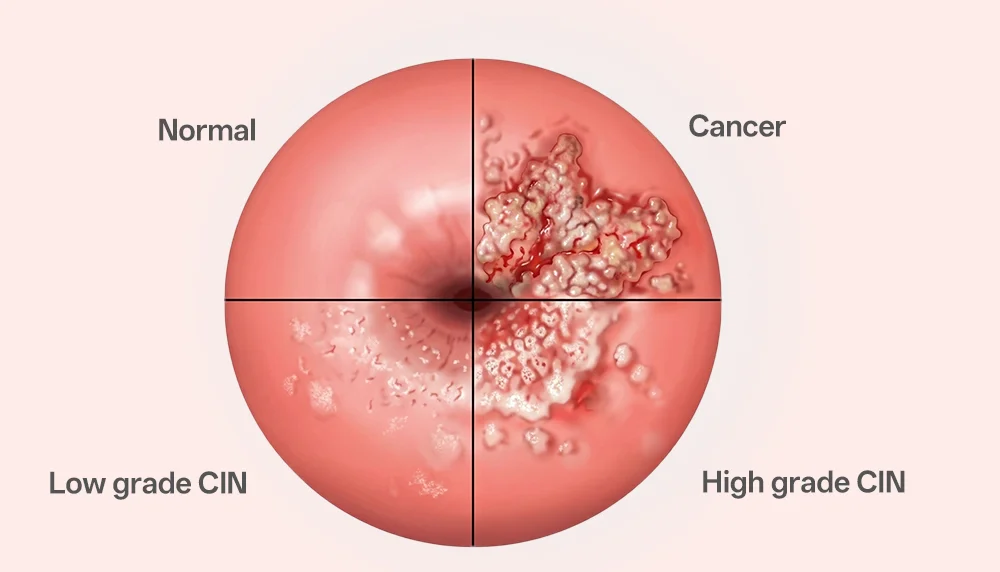
There is no reliable appearance chart that can assign a cervical cancer stage. Staging describes how far cancer has spread. It is based on examination, biopsy, imaging, and other tests.
The table below explains the general pattern without suggesting that stage can be read from the surface of the cervix.
| Stage | What the stage means | What may be noticed |
|---|---|---|
| Stage IA | Cancer is very small and can be identified only under a microscope | The cervix may look normal. There may be no visible lesion. |
| Stage IB | Cancer is larger but still limited to the cervix | A lesion may become visible as it grows. It can be friable, raised, ulcerated, or irregular, but appearance varies. |
| Stage II | Cancer has spread beyond the cervix and uterus, but not to the pelvic wall or lower third of the vagina | An exam may suggest extension into the upper vagina or tissues beside the cervix. Imaging and a full assessment are needed to define the extent. |
| Stage III | Cancer involves the lower vagina, pelvic wall, kidneys, or certain lymph nodes | There may be a larger fixed pelvic lesion or signs caused by urinary obstruction. The cervix alone cannot show the full stage. |
| Stage IV | Cancer has reached the bladder, rectum, or distant organs | Local tissue damage may be extensive, but scans, biopsy, and other tests are needed to identify organ or distant spread. |
Early cervical cancer may be invisible. A dramatic-looking cervical lesion is not automatically late-stage cancer. Conversely, a cervix that looks normal does not replace recommended screening.
When Does an Abnormal Cervix Need Follow-Up?
Follow-up depends on the appearance, symptoms, and screening history. Prompt assessment is especially important for:
- bleeding after sex, between periods, or after menopause;
- a visible ulcer, mass, or irregular lesion;
- a cervix that bleeds easily without a clear benign cause;
- persistent watery, bloody, or foul-smelling discharge;
- unexplained pelvic pain;
- an abnormal Pap or positive high-risk HPV result;
- an abnormal-looking cervix even when screening is normal.
The clinician may take swabs, repeat or arrange screening, refer for colposcopy, or take a biopsy. The safest next step is based on the whole picture, not one visual feature.
Conclusion
An abnormal cervix appearance does not always mean cancer. Ectropion, cervicitis, cervical polyps, and nabothian cysts are common examples of benign or treatable findings.
Some appearances, such as a white plaque, an ulcer, a hard nodule, or a friable irregular mass, need closer assessment. The key is to match the visual examination with the right test. Pap and HPV tests support screening, colposcopy provides a closer view, and biopsy confirms the tissue diagnosis.
Cervical cancer stage cannot be determined by appearance alone. In fact, the earliest stage may not be visible at all.
Frequently Asked Questions
Can a cervix look abnormal even when a Pap test is normal?
Yes. A Pap test samples cells from part of the cervix. A polyp, cyst, inflamed area, or visible lesion may still need assessment even if cytology is normal.
Does bleeding during a cervical exam mean cancer?
No. Ectropion, cervicitis, a polyp, pregnancy-related changes, and recent irritation can all cause contact bleeding. Unexplained or repeated bleeding still needs evaluation so more serious causes are not missed.
Can a clinician tell the difference between cervicitis and cervical cancer by looking?
Not with certainty. Both can cause redness, discharge, irregular tissue, or easy bleeding. Infection tests, colposcopy, and biopsy may be needed when the cause is unclear or the appearance is suspicious.
Sources and Medical Disclaimer
- National Cancer Institute: Cervical Cancer Diagnosis
- National Cancer Institute: Cervical Cancer Stages
- National University Health System: Cervical Cancer
- Australian Centre for the Prevention of Cervical Cancer: Key Information for Health Professionals
- American College of Obstetricians and Gynecologists: Colposcopy
- CDC: Urethritis and Cervicitis Treatment Guidelines
This article is for medical education only. It is not a diagnosis or a substitute for patient-specific advice, cervical screening guidance, colposcopy, pathology, or specialist care. Screening and referral pathways vary by country and clinical setting.
Training Resources
The Pap Smear Test Model supports supervised practice in speculum insertion, cervical visualization, and cervical cell sampling. More teaching options are available in the Women’s and Reproductive Health Training Models collection.