Granulation tissue is the new, vascular tissue that fills an open wound as it heals. Healthy granulation is usually a welcome sign, but color alone cannot tell you whether a wound is progressing normally. Red tissue may be healthy granulation, fragile hypergranulation, inflammation, or another abnormal change; yellow or black tissue may represent slough or eschar and requires a different assessment.
This guide explains what granulation tissue is, what healthy granulation looks like, how it differs from hypergranulation, slough, eschar, and epithelial tissue, and when a wound needs prompt clinical review.
Important: This article is for wound-care education and clinical training. It does not diagnose a wound or replace assessment by a licensed healthcare professional. Follow the patient’s care plan and local wound-care protocol.
What Is Granulation Tissue?
Granulation tissue is temporary repair tissue that develops mainly during the proliferative phase of wound healing. It contains new capillaries, fibroblasts, extracellular matrix, collagen, and immune cells. Together, these components help fill a wound from the base, restore blood supply, support contraction, and provide a surface over which epithelial cells can migrate.
The word granulation describes the tissue’s fine, grain-like appearance. It is not related to a granuloma, which is a different inflammatory process.
Granulation tissue is especially visible in wounds healing by secondary intention—wounds whose edges are not brought together and that must fill from the bottom upward. It may also be seen in chronic wounds, pressure injuries, traumatic wounds, surgical wounds left open, and around stomas or tubes.
Where Granulation Fits in the Wound-Healing Process
Wound healing is often described in four overlapping phases:
- Hemostasis: vessels constrict, platelets aggregate, and a clot helps stop bleeding.
- Inflammation: immune cells remove bacteria, damaged tissue, and debris while signaling the next phase of repair.
- Proliferation: fibroblasts build matrix and collagen, new vessels grow, granulation tissue fills the defect, and epithelial cells begin covering the surface.
- Remodeling: collagen is reorganized and the repaired tissue gradually gains strength.
Granulation is therefore evidence that repair activity is occurring, but it does not mean the wound is closed or that the underlying cause has been corrected. A red wound bed can remain stalled when pressure, poor perfusion, infection, friction, excess moisture, or another barrier persists.
What Does Healthy Granulation Tissue Look Like?
Healthy granulation tissue is commonly:
- Pink-red to bright red because it contains many new capillaries.
- Moist and slightly glossy, without being waterlogged.
- Finely bumpy or pebbled rather than smooth and leathery.
- Level with or slightly below the wound edge, allowing epithelial cells to migrate across it.
- Delicate, so a small amount of spotting may occur when it is touched during care.
The finding should be interpreted as part of a trend. Healthy progress usually includes decreasing wound dimensions, advancing epithelial edges, manageable exudate, and stable or improving pain. A wound that remains the same size—or becomes larger, deeper, more painful, or more heavily draining—needs reassessment even if part of the bed looks red.
What Does a Granulating Wound Bed Look Like Day by Day?
A granulating wound is one whose base is actively filling with this new red repair tissue, and its appearance changes as healing advances rather than staying the same from day to day. In an open wound healing from the base upward, the granulating wound bed usually moves through a recognizable sequence.
- Early: the base is still shallow and may show a mix of red granulation with residual slough or fibrin, so only part of the bed looks red.
- Progressing: healthy granulation spreads across more of the bed, becomes more uniformly pink-red and finely pebbled, and rises to sit level with the surrounding tissue.
- Approaching closure: the bed is filled to the level of the edges, and pale pink or pearly epithelial tissue begins advancing inward from the margins or as small islands.
The useful signal is the direction of change, not a single day’s snapshot. A granulating wound bed that keeps filling, contracting, and epithelializing is progressing; one that stalls at the same appearance, stops contracting, or becomes increasingly friable should be reassessed even when the tissue still looks red.
Granulation Tissue vs Epithelial Tissue
Granulation and epithelial tissue are both involved in healing, but they play different roles.
| Tissue | Typical appearance | Role in healing |
|---|---|---|
| Granulation tissue | Moist, pink-red or red, finely bumpy tissue in the wound bed | Fills the tissue defect and supports repair from the base |
| Epithelial tissue | Thin, pale pink or pearly tissue advancing from the edges or appearing as small islands | Covers the wound surface with new skin |
A healthy red wound bed is not yet a closed wound. Newly epithelialized tissue is also fragile and continues to mature during remodeling.
Healthy Granulation vs Hypergranulation
Hypergranulation—also called overgranulation or proud flesh—is granulation tissue that rises above the level of the surrounding skin. Because epithelial cells migrate across a relatively level surface, this raised tissue can delay surface closure.
Hypergranulation may appear:
- Bright red, dark red, or purple-red.
- Shiny, soft, spongy, or jelly-like.
- Raised or nodular rather than level with the wound edge.
- Friable — easily irritated and prone to bleeding when touched.
- Associated with persistent moisture or drainage.
Possible contributors include excessive moisture, friction or movement, foreign material, bacterial burden, local infection, and prolonged inflammation. Tubes, devices, or dressings that repeatedly rub the same area can be important clues.
Management should address the cause rather than simply remove visible tissue. A clinician may reassess moisture control, dressing choice, pressure or friction, device stability, foreign material, and signs of infection. Topical treatments or procedures should only be used under an appropriate clinical plan. Patients should not cut, burn, or apply caustic home remedies to raised tissue.
Granulation Tissue vs Slough and Eschar
Granulation, slough, and eschar can be relevant when assessing full-thickness pressure injuries, but they are not a fixed sequence through which every pressure injury progresses. Pressure injury stages are assigned according to the deepest tissue type exposed, not by wound-bed color alone, and a healing injury is not reverse staged. Stage 2 does not contain granulation tissue, slough, or eschar. Granulation and slough may be visible in Stage 3; Stage 4 is defined by exposed or directly palpable deeper structures, while slough or eschar may also be present. If slough or eschar obscures the depth of tissue loss, the injury is unstageable until the extent can be determined.
| Finding | Common appearance | Key distinction |
|---|---|---|
| Healthy granulation | Pink-red or bright red; moist; pebbled | Vascular repair tissue |
| Hypergranulation | Red to purple-red; soft or shiny; raised above the edge | Excess repair tissue that may block epithelialization |
| Slough | Yellow, cream, tan, gray, or sometimes green; moist, soft, stringy, or gelatinous | Devitalized tissue or debris attached to the wound bed |
| Eschar | Brown or black; dry, thick, firm, or leathery | Devitalized tissue that forms a covering over the wound |
For pressure-injury training, this distinction matters because learners should describe the wound bed before assigning a stage: identify viable granulation, estimate any slough or eschar, determine whether deeper structures are exposed, and recognize when obscured depth prevents staging.
Do not identify tissue from color alone. Yellow material can include slough, fibrin, exudate, or other debris; black material may be eschar, dried blood, or another change. The clinician must consider wound cause, perfusion, location, depth, moisture, attachment, and the patient’s overall condition.
Debridement decisions are wound-specific. Slough or eschar may need clinician-directed removal to expose the wound bed and support healing, but there are important exceptions. For example, dry, stable eschar on an ischemic foot or heel may require vascular or specialist assessment before any attempt to remove it. Do not debride tissue without the appropriate training, authorization, and clinical plan.
Granulation Tissue vs Wound Infection
Granulation tissue itself is not pus and does not automatically mean infection. Conversely, the presence of red tissue does not rule infection out. Look for a pattern of change across the wound and the patient.
| Feature | More consistent with healthy progress | Infection or deterioration concern |
|---|---|---|
| Pain | Stable or improving | New, increasing, or disproportionate pain |
| Periwound | Stable skin without spreading change | Increasing heat, swelling, redness, or discoloration |
| Drainage | Stable amount expected for the wound | New purulent drainage, sudden increase, or unpleasant odor |
| Wound bed | Moist red tissue with improving dimensions | Dusky, gray, increasingly friable, or rapidly changing tissue |
| General condition | No systemic symptoms | Fever, chills, malaise, confusion, or feeling acutely unwell |
Redness can be harder to recognize in brown or black skin. Compare the area with surrounding skin and assess temperature, swelling, firmness, pain, and color change rather than relying on visible redness alone.
Suspected infection requires timely clinical review. A dressing change by itself is not a substitute for assessing the wound, the patient, and the need for further investigation or treatment.
Assess the Whole Wound, Not Just Its Color
A repeatable assessment helps prevent a single visual feature from driving the conclusion. Document:
- Cause and location: pressure, vascular, neuropathic, surgical, traumatic, moisture-associated, or another cause.
- Dimensions: length, width, depth, undermining, and tunneling, using the same method each time.
- Wound-bed tissue: estimated proportions of granulation, epithelial tissue, slough, eschar, and any exposed structures.
- Exudate: amount, color, consistency, and odor after cleansing.
- Edges: attached, advancing, rolled, macerated, or undermined.
- Periwound skin: heat, swelling, discoloration, maceration, dryness, or breakdown.
- Pain and function: baseline, change over time, and pain during care.
- Systemic factors: perfusion, pressure exposure, glucose control, nutrition, medications, mobility, and infection risk.
Use photographs only according to consent, privacy, and facility policy. Measurements and descriptions are most useful when recorded consistently so that the team can compare trends rather than isolated snapshots.
Principles That Support Healthy Granulation
The exact care plan depends on the wound’s cause, depth, perfusion, drainage, infection risk, and the patient’s health. Broad clinical principles include:
- Correct the cause: offload pressure, reduce repeated trauma, manage shear, and stabilize rubbing tubes or devices.
- Maintain moisture balance: protect viable tissue from drying while preventing leakage and periwound maceration.
- Cleanse according to protocol: use the prescribed method and avoid harsh or unapproved substances.
- Protect wound edges: select dressings and skin protection appropriate to the amount of exudate.
- Address devitalized tissue safely: make debridement decisions according to wound type, perfusion, risk, and scope of practice.
- Reassess unexpected change: increasing pain, drainage, odor, discoloration, or stalled dimensions should trigger review.
- Support the whole patient: circulation, nutrition, glucose management, mobility, pressure redistribution, smoking status, and adherence all influence healing.
The goal is not simply to produce more red tissue. It is to create the conditions for orderly granulation, epithelial advancement, wound contraction, and eventual remodeling.
When to Seek Prompt Medical Review
Arrange prompt clinical assessment for:
- Spreading redness or discoloration, increasing warmth, swelling, or pain.
- Pus, a new unpleasant odor, a sudden increase in drainage, or wound separation.
- Fever, chills, confusion, red streaking, or feeling generally unwell.
- Tissue that becomes gray, black, rapidly darker, or unusually friable.
- Bleeding that is heavy or does not stop with appropriate gentle pressure.
- A wound that becomes larger, deeper, or fails to progress.
- Raised tissue that blocks closure or repeatedly bleeds.
- Any concerning wound in a person with diabetes, poor circulation, reduced sensation, or immune suppression.
Urgency depends on the patient’s condition and local pathway. Severe pain, rapidly spreading change, systemic illness, or concern about compromised blood flow warrants urgent evaluation.
Conclusion
Healthy granulation tissue is typically moist, pink-red to bright red, finely bumpy, and contained within the wound bed. It supports closure, but it is only one part of the healing picture. Hypergranulation rises above the wound edge and may delay epithelialization; slough and eschar are devitalized tissues with different assessment needs; infection is judged from the pattern of local and systemic changes, not from color alone.
The safest approach is structured, repeated assessment: describe what is visible, measure the trend, consider the wound’s cause and the whole patient, and escalate unexpected change.
Frequently Asked Questions
How long does granulation tissue stay in a wound?
There is no fixed duration. Granulation may be visible for days or weeks while an open wound fills and epithelial tissue advances. Wound size, depth, blood flow, pressure, infection, underlying disease, and treatment all affect the timeline. A wound that is not measurably progressing should be reassessed rather than judged by elapsed time alone.
Should granulation tissue be covered with a dressing?
Open granulation tissue is generally protected with a dressing chosen for the wound’s depth, drainage, location, and care plan. The aim is balanced moisture and protection from trauma—not leaving the tissue dry or allowing excess fluid to macerate the surrounding skin. Dressing selection should follow clinician guidance and local protocol.
Does granulation tissue turn directly into new skin?
Not exactly. Granulation tissue fills and supports the wound bed. Epithelial cells then migrate across that surface to create a new skin covering, while deeper tissue continues to remodel. Both processes contribute to closure, but they are different tissue stages.
What does friable granulation tissue mean?
Friable means the tissue is fragile and bleeds easily when touched, even during a gentle dressing change. A small amount of spotting from healthy granulation can be normal, but tissue that is increasingly friable — bleeding readily, looking dusky, or breaking down — can point to hypergranulation, a high bacterial burden, or another problem, and warrants clinical review rather than reassurance from red color alone.
Sources and Medical Disclaimer
- NCBI Bookshelf: Physiology, Granulation Tissue
- NCBI Bookshelf: Wound Healing Phases
- NHS Scotland Right Decisions: Visual Wound Guide
- University Hospitals Sussex NHS Foundation Trust: Hypergranulation
- NCBI Bookshelf: Nursing Fundamentals—Slough and Eschar
- Guy’s and St Thomas’ NHS Foundation Trust: Signs of Wound Infection
This content is educational and is not a substitute for diagnosis, treatment, or an individualized wound-care plan from a qualified healthcare professional.
Training Resources
Accurate tissue recognition improves when learners can compare wound features repeatedly before patient contact. The Pressure Ulcer Model 4 Stages Kit can support a structured observation exercise: compare intact non-blanchable erythema, partial-thickness loss, full-thickness loss with granulation or slough, and deeper injury while asking learners to distinguish pressure-injury stage from wound-bed tissue. The model is a teaching aid; it does not imply that every pressure injury passes through these appearances in order, and an actual wound may be unstageable when slough or eschar obscures its depth. Educators building a broader skills lab can also explore Wound Care, Ulcer & Stoma Models for practice with wound description, staging, dressing decisions, and documentation.
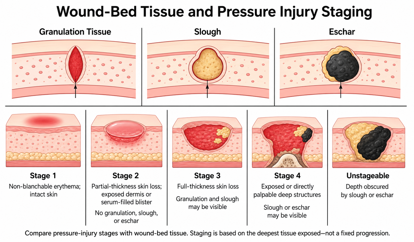
Wound-bed tissue and pressure injury staging: categories for comparison, not a fixed progression.