If you draw blood from dogs competently, most of that skill transfers to cats. The problem is the part that does not, because it is not obvious which part that is.
Nothing about the puncture itself is different. What changes is the set of defaults you bring to the table. Which vein you reach for first. How much blood you can plan on. What equipment you pick up. And how much the patient’s tolerance drives the decision.
This article is the translation table. It does not walk through the procedure for either species — those are covered separately for dogs and for cats — and it does not cover handling technique, which is in the low-stress restraint guide.
Quick Answer: The Four Defaults That Flip
| Dog | Cat | |
|---|---|---|
| Routine hind limb site | Lateral saphenous, outer surface | Medial saphenous, inner surface |
| Role of the jugular | Reserved for larger volumes | Moves up the list; often the practical choice |
| Volume headroom | Usually generous for routine panels | Tight enough to change the sample plan |
| What drives site choice | Vessel and sample requirements | Vessel, sample, and what the patient will tolerate |
Everything else — aseptic preparation, needle handling, sample care, knowing when to stop — carries over unchanged.
The Hind Limb Site Is on the Other Side
This is the one that marks out a handler who has not worked with cats.
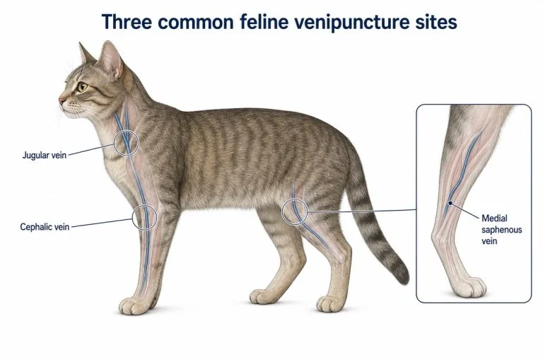
In dogs, the routine hind limb vessel is the lateral saphenous, on the outer surface of the leg. In cats, it is the medial saphenous, on the inner surface. They are different vessels, approached from opposite sides, and a technique carried straight across from dogs will be looking at the wrong side of the limb.
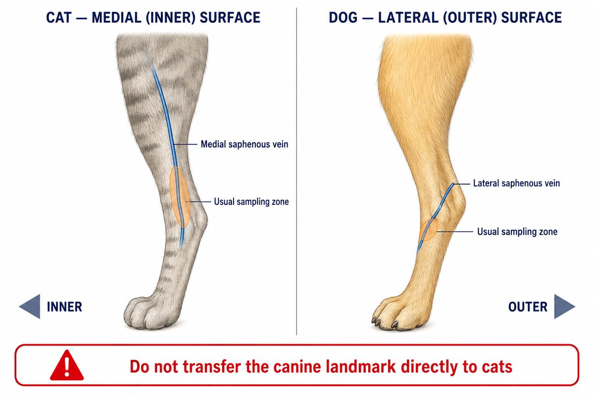
The routine hind limb vessel sits on opposite surfaces in the two species. Orange marks the usual sampling zone: the medial tibia in the cat, just above the hock in the dog. Joint angulation is simplified to keep the vessel course readable.
The reason is practical rather than anatomical. In cats the medial saphenous is the larger and more accessible of the two. It is also reached with the patient lying on its side, which most cats tolerate far better than neck handling.
The Jugular Changes Rank
In dogs, the jugular is generally where you go when a peripheral vein will not deliver the volume you need. It is a step up rather than a starting point.
In cats, it moves up the list. The peripheral vessels are small enough that filling several tubes from a limb is slow, and a slow collection in a small vein is where samples clot and get damaged. For a full panel on a cat, the jugular is frequently the sensible first choice rather than the fallback.
The trade-off is that neck handling is what many cats object to most. That is why the decision is not purely a vessel decision in cats — see below.
Volume Arithmetic Stops Being Comfortable
The safety rule is the same for both species. A healthy patient can give roughly one percent of body weight. The maximum single draw is around ten percent of total blood volume, with a recovery interval before the next one.
What changes is the headroom that rule leaves you.
For a 30 kg dog, a routine panel is a small fraction of what is available, and the calculation rarely constrains anything. For a 5 kg cat the same rule leaves a few milliliters of working room, and that is before you account for a patient who may be anemic, dehydrated, or small for its age.
So in cats the sample plan has to be made before the needle comes out. Which tests actually matter today. Which tubes they need. Whether smaller-volume tubes are appropriate. And what you will drop if the collection runs short. In dogs that planning is usually optional. In cats it decides whether the patient gets stuck twice.
Feline volume limits and the site-by-volume decision are covered in detail in the cat blood draw sites guide.
Your Equipment Defaults Change Too
Two habits carried over from dogs cause avoidable failures in cats.
Too much suction. A syringe that is comfortably sized for a canine draw applies more negative pressure than a feline vein can tolerate. The vessel collapses around the needle, flow stops, and the natural response — pulling harder — makes it worse. Use a smaller syringe, or a butterfly set, and draw gently.
Needle and system choice. A smaller vessel and a slower fill favor a system you can control with one hand and hold still for longer. The needle that works on a dog’s cephalic is not automatically the right choice on a cat’s.
Neither of these is a technique problem. Both are equipment defaults that need resetting.
Patient Tolerance Becomes a Clinical Input
In dogs, how well the patient copes rarely changes which vein you use. In cats it often does, and treating it as a soft consideration is how a first attempt becomes a third.
A cat that will not accept neck handling but settles on its side has effectively made the site decision for you: the medial saphenous, not the jugular, even if the jugular would fill the tubes faster. Forcing the theoretically better site on a patient that will not hold still costs you three things: a damaged sample, a bruised vessel, and a harder patient next visit.
This is a decision input, not a handling technique. How to hold and position a cat is covered in the low-stress restraint guide.
What Does Not Change
It is worth being explicit about this, because a list of differences can make cats sound like an unrelated procedure. They are not.
Aseptic preparation is the same. Anchoring the vessel and entering at an angle appropriate to a superficial vein is the same. Filling tubes in the correct order, mixing them properly, and getting them to the laboratory promptly is the same. Applying pressure afterwards is the same. Recognizing that repeated attempts damage the vessel and the patient, and stopping before that point, is the same.
If you are competent with dogs, you are not starting over. You are resetting four defaults.
Conclusion
Moving from dogs to cats is not a matter of doing the same procedure more delicately. It is a matter of changing what you reach for first.
The hind limb site is on the inner surface, not the outer. The jugular is a realistic first choice rather than an escalation. The volume rule leaves you far less room, so the sample has to be planned in advance. And what the patient will tolerate is part of the clinical decision rather than an inconvenience around it.
Everything else you already know still applies.
Frequently Asked Questions
Why is the saphenous vein on a different side in cats?
The vessel that is routinely used is different, not the anatomy of one vein. Dogs are typically sampled from the lateral saphenous, on the outer surface of the hind limb. Cats are typically sampled from the medial saphenous, on the inner surface. In cats that vessel is the larger and more accessible of the two, and it is reached in a position most cats tolerate well.
Can you take the same volume of blood from a cat as from a dog?
Not in absolute terms. The percentage-based safety rule is the same for both, but a cat’s much smaller total blood volume means the same rule leaves only a few milliliters of working room. Plan which tests matter before collection, and account for patients who are anemic, dehydrated, or unusually small.
Do I need different equipment for cats?
Often yes, and the reason is suction rather than size alone. A syringe sized for a canine draw applies more negative pressure than a small feline vein tolerates, which collapses the vessel and stops the flow. A smaller syringe or a butterfly set, drawn gently, is usually the better default.
Sources
- Merck Veterinary Manual — Collection and Submission of Laboratory Samples from Animals (merckvetmanual.com)
- Today’s Veterinary Nurse — Uncommon IV Catheter Sites in Small Animals (todaysveterinarynurse.com)
- American Association of Feline Practitioners / International Society of Feline Medicine — 2022 Cat Friendly Veterinary Interaction Guidelines (catvets.com)
- dvm360 — Tips for technicians to improve phlebotomy skills (dvm360.com)
Disclaimer: This article is for professional education only and is not a substitute for veterinary advice, diagnosis, or treatment. Sample volumes, site selection, and handling decisions for an individual patient must follow your practice’s protocols and the direction of the attending veterinarian. Last updated: September 2026.
Training Resources
Resetting a default is easier when the hand sequence underneath it is automatic, and that part is best rehearsed away from a patient.
The Veterinary Cat IV Practice Kit is a feline peripheral-vein leg model for practicing vein puncture, blood draw with realistic flashback, IV catheter placement, and fluid-infusion setup. It represents a limb vein, so it covers peripheral practice only; it is not a jugular model.
For the canine side, the Dog IV Practice Kit is a full dog leg IV catheter kit, and the Canine IV Training Model is the basic veterinary IV catheter practice kit covering the same core functions with fewer accessories.
Related guides: feline IV catheter placement and canine IV catheterization.