Pressure ulcers — also called bedsores or pressure injuries — are localized damage to the skin and underlying tissue caused by sustained pressure over a bony prominence. For clinical staff, the work comes down to three things this guide focuses on: assessing risk, staging accurately, and matching the right treatment to each stage. Getting the stage right matters because it drives the entire care plan — and, as any wound nurse knows, a few classic pitfalls trip clinicians up again and again.
This is the clinical, staging-and-treatment piece of our pressure injury series. For the other angles, see our companion guides: spotting pressure injuries early (including on darker skin), how long pressure ulcers take to heal, and preventing bedsores at home.
Risk Assessment: the Braden Scale
Prevention starts with identifying risk early, and the Braden Scale is the validated tool most units rely on. It scores six factors — sensory perception, moisture, activity, mobility, nutrition, and friction/shear. Lower totals mean higher risk: a score of 18 or below generally flags risk, with progressively more intensive prevention as scores fall into the high-risk (≤12) range. Reassess on admission, after any change in condition, and at routine intervals, and trigger a prevention bundle the moment a patient screens at-risk. (The full at-home prevention routine — the SSKIN bundle — is covered in our prevention guide.)
The Pressure Ulcer Stages, and the Care Each Demands
Pressure ulcers are classified by the depth of tissue damage, following the international NPIAP/EPUAP system. Accurate staging drives the care plan — including dressing choice — so it is worth knowing each category precisely.
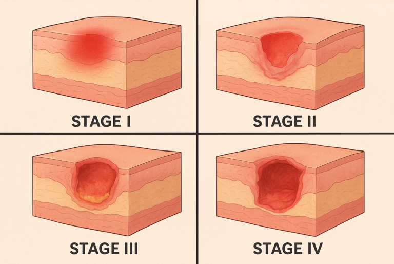
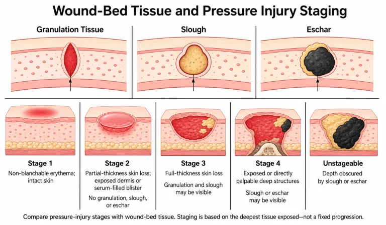
Stage 1: Non-blanchable redness, intact skin
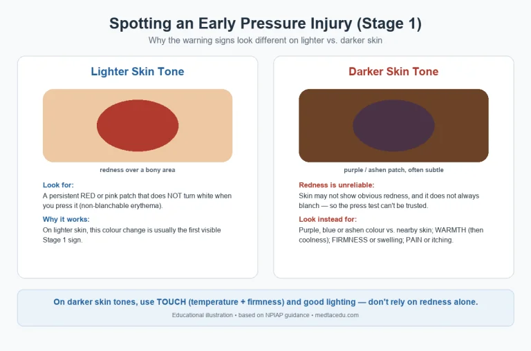
The skin is unbroken but shows redness that does not blanch when pressed (on darker skin tones it may look purple or blue and feel warmer or firmer — see our early-signs guide for assessing darker skin). Nursing care: offload immediately, reposition at least every two hours, and protect the skin with a transparent film or barrier film if needed — no absorbent dressing required. Fully reversible if pressure is relieved promptly.
Stage 2: Partial-thickness skin loss
The epidermis is broken, exposing the dermis — a shallow open ulcer with a pink-red bed, or an intact/ruptured blister. Nursing care: maintain a clean, moist wound environment with a hydrocolloid or thin foam dressing, manage moisture, continue offloading, and monitor for infection.
Stage 3: Full-thickness skin loss
Damage extends through the skin into subcutaneous fat — a deeper crater, slough may be present, but bone/tendon/muscle are not exposed. Nursing care: wound bed preparation is central — debride dead tissue as ordered, fill dead space with an alginate or foam dressing (hydrogel if the bed is dry), optimise nutrition, and monitor for infection.
Stage 4: Full-thickness loss with exposed structures
The deepest category: tissue loss exposes bone, tendon, or muscle, with a high risk of osteomyelitis and systemic infection. Nursing care: a coordinated, multidisciplinary plan — advanced wound management (frequently NPWT), possible surgical debridement or flap closure, aggressive infection control, pain management, and nutritional support.
⭐ Common Staging Pitfalls (Where Clinicians Go Wrong)
Accurate staging is a skill, and a handful of mistakes show up over and over. Avoiding these is what separates confident wound assessment from guesswork.
1. Mistaking a Deep Tissue Injury for Stage 2
This is the most consequential error. A Stage 2 is a shallow, partial-thickness wound — a blister or abrasion with a pink-red bed. A Deep Tissue Injury (DTI) is intact or blistered skin showing persistent deep purple or maroon discoloration, with serious damage developing beneath the surface. Labelling a DTI as “just a Stage 2” badly underestimates it — DTIs can open into full-thickness wounds within days. If the discoloration is deep purple/maroon rather than pink-red, think DTI, not Stage 2.
2. Trying to stage an Unstageable ulcer
When the wound base is obscured by slough or eschar, the true depth cannot be seen — so it is Unstageable, not “Stage 3” or “Stage 4” by guesswork. It can only be staged once enough non-viable tissue is removed to reveal the wound bed. (One exception: stable, dry, intact eschar on an ischaemic heel is often left in place rather than debrided.)
3. Confusing moisture damage (MASD/IAD) with a pressure injury
Incontinence-associated dermatitis (IAD) and other moisture-associated skin damage (MASD) are frequently miscoded as Stage 1 or 2 pressure injuries. The tells: moisture damage is usually diffuse with irregular edges, sits in skin folds or the perineal/peri-anal area, and is linked to wet skin — not necessarily over a bony prominence. A pressure injury is typically over a bony prominence, more localized with defined edges, and linked to pressure/shear. Getting this distinction right changes the whole care plan (barrier protection and moisture management vs. offloading).
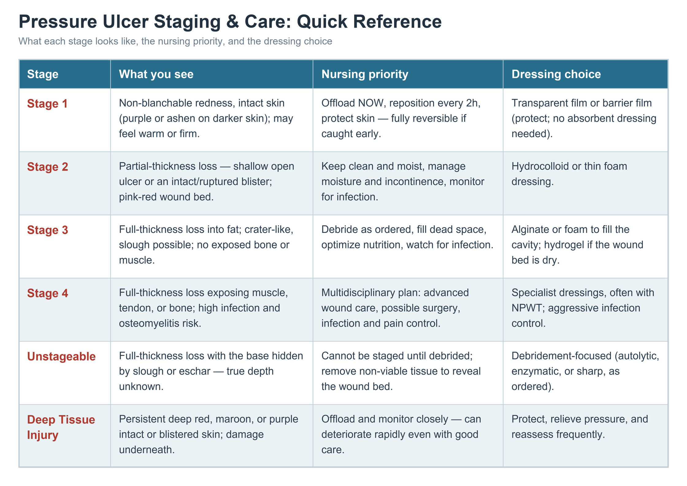
Quick reference: staging, priority, and dressing at a glance
Stage-Specific Treatment: Dressings, Debridement & NPWT
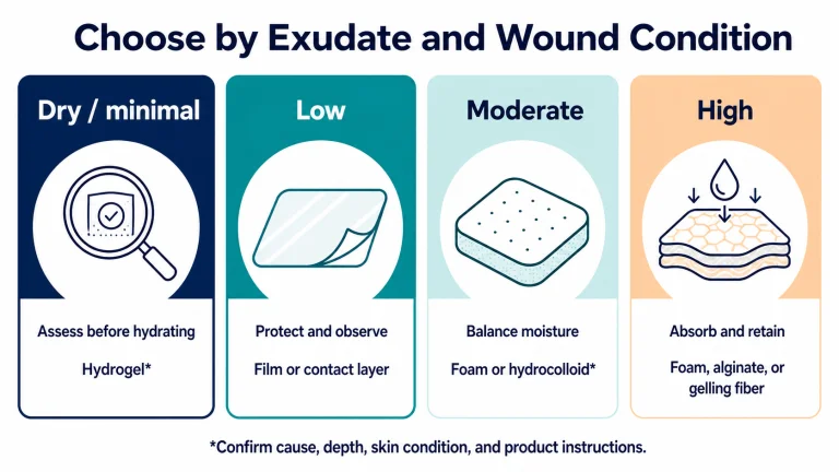
Dressings follow the stage and the wound’s moisture balance, with one unifying principle — keep the wound bed moist while managing exudate: film/barrier film for Stage 1, hydrocolloid or foam for Stage 2, alginate or foam (hydrogel if dry) to fill Stage 3 cavities, and specialist dressings for Stage 4.
Debridement removes non-viable tissue so healthy tissue can rebuild. The main methods, chosen by wound and patient factors, are: autolytic (moisture-retentive dressings let the body’s own enzymes self-digest slough — selective but slow), enzymatic (a topical agent digests dead tissue), mechanical (e.g., wet-to-dry or monofilament pads — non-selective), and sharp/surgical (fastest, performed by a trained clinician). Debridement is a prerequisite before staging an unstageable wound or starting NPWT.
Negative Pressure Wound Therapy (NPWT), or vacuum-assisted closure (VAC), is used for deeper Stage 3–4 wounds after adequate debridement. A foam dressing sealed with an airtight film delivers controlled suction that draws off exudate and infectious material, reduces swelling, helps pull wound edges together, and stimulates blood flow and granulation tissue — often a cleaner bed and faster healing than conventional dressings. It is applied and monitored by a wound specialist, with dressings changed every two to three days, and is most valuable for large, deep, or heavily exuding ulcers that are struggling to progress.
Wound Assessment & Documentation
Consistent documentation is what proves a wound is progressing — or flags early that it is not. At each assessment, record: location and stage; size (length × width × depth, plus any undermining or tunnelling); wound bed (granulation, slough, eschar, percentages); exudate (amount, type, odour); wound edges and surrounding skin; and pain. Photograph per facility policy, measure consistently the same way each time, and track the trend visit to visit. A wound that is not measurably improving within an expected window — or that shows signs of infection (spreading redness, warmth, odour, purulent drainage, fever) — should be escalated.
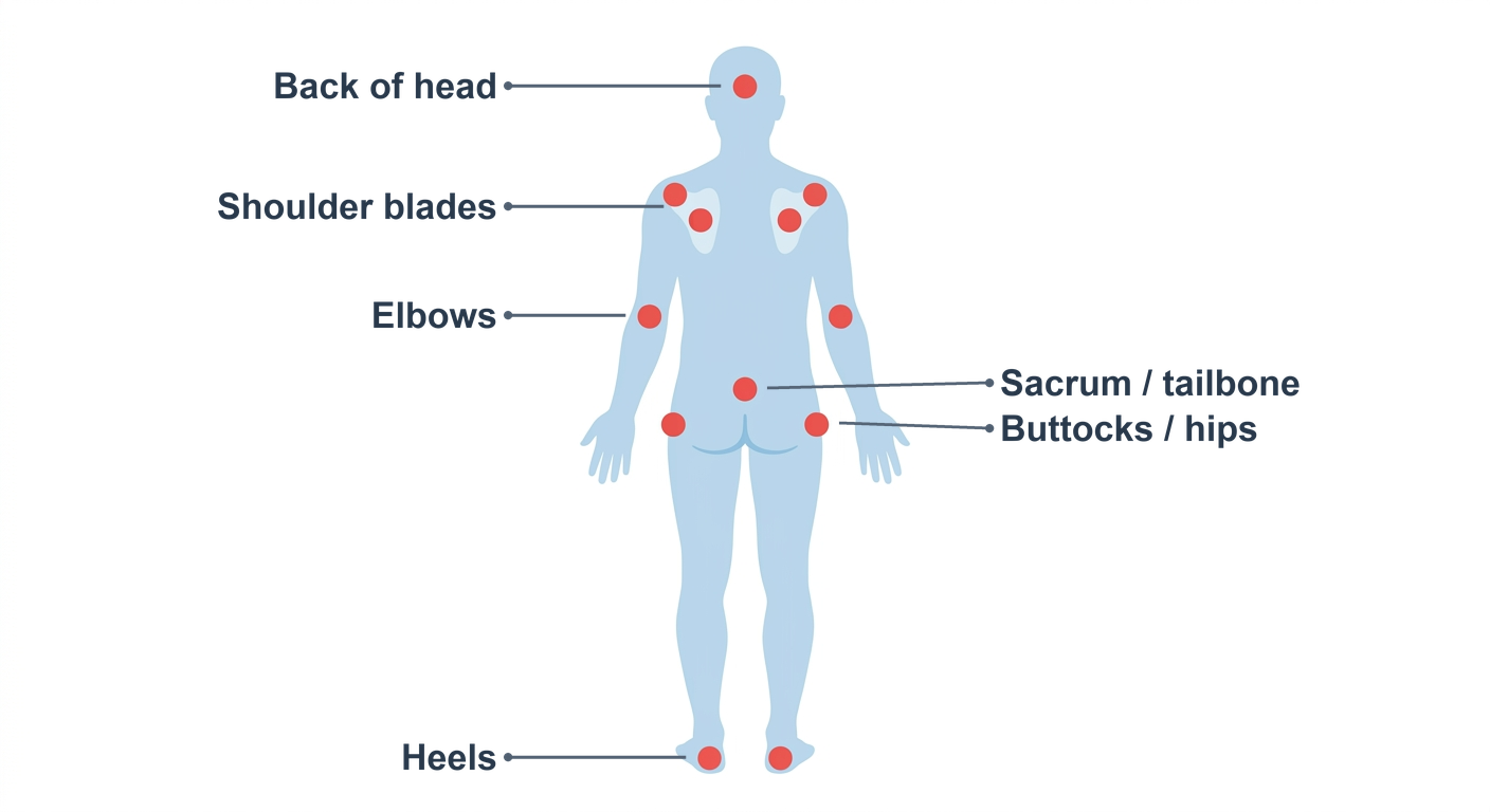
Where Pressure Ulcers Form — Inspect These First
Pressure injuries cluster over bony prominences. During every skin inspection, prioritise the sacrum, coccyx, and heels (supine patients), the hips/greater trochanter and outer ankles (side-lying), and the ischial tuberosities (“sit bones”) for seated patients. Don’t miss device-related sites: behind the ears (oxygen tubing), under catheters, collars, and casts. Inspection has to follow the patient’s posture, not a fixed checklist — the figure below maps the high-risk sites at a glance (full prevention detail is in our prevention guide).
Nursing Care Plan for Pressure Ulcers
A structured nursing care plan turns staging and assessment into consistent, measurable care. Most pressure-ulcer (pressure-injury) care plans follow the standard nursing-process framework:
1. Nursing diagnoses (common examples)
- Impaired skin integrity related to pressure, shear, or moisture
- Risk for infection related to compromised skin and an open wound
- Imbalanced nutrition: less than body requirements
- Impaired physical mobility
- Acute or chronic pain related to tissue damage
2. Goals and expected outcomes (make them measurable)
- The wound shows signs of healing — reduced size and granulation — within a set timeframe
- No signs of infection (no spreading redness, purulent drainage, or fever)
- The patient or caregiver can describe pressure-relief and skin-care measures
- Skin over remaining at-risk areas stays intact
3. Nursing interventions
- Pressure redistribution: reposition on an individualized schedule, use appropriate support surfaces, and offload the heels
- Skin care: keep skin clean and dry, manage moisture and incontinence, and inspect at-risk areas each shift
- Wound care: stage-appropriate cleansing and dressings, debridement as ordered, and monitoring for infection
- Nutrition: ensure adequate protein, calories, and hydration; involve a dietitian for higher-stage or non-healing wounds
- Education: teach the patient and family repositioning, skin checks, and the warning signs to report
4. Evaluation
Reassess the wound and the plan regularly: measure the wound, document healing or deterioration, and revise the interventions whenever the goals aren’t being met. This care-plan structure pairs directly with the staging and treatment sections above — stage the wound accurately, then build the plan around the patient’s risk factors and the wound’s stage.
When to Escalate
Notify the wound care team or physician when an ulcer advances a stage, shows signs of infection, fails to progress toward healing, or whenever a Stage 3, Stage 4, unstageable, or deep tissue injury is identified. Early escalation prevents a manageable wound from becoming a life-threatening one.
Frequently Asked Questions
How do I tell a Stage 2 from a deep tissue injury?
Colour and depth. A Stage 2 is a shallow, partial-thickness wound — a blister or abrasion with a pink-red bed. A deep tissue injury is intact or blistered skin with persistent deep purple or maroon discoloration and damage developing underneath. Deep purple/maroon = think DTI, not Stage 2.
Why can’t an unstageable ulcer be staged?
Because slough or eschar covers the wound base, so you cannot see how deep the damage goes. Once enough non-viable tissue is debrided to reveal the bed, it is staged (usually Stage 3 or 4).
How often should a pressure ulcer dressing be changed?
It depends on the dressing and exudate. Hydrocolloids and foams are often left several days (up to ~5–7) if intact and not saturated; heavily exuding or infected wounds need more frequent changes; NPWT dressings are typically changed every 2–3 days. Always follow the wound care plan and change sooner if a dressing leaks, loosens, or infection is suspected.
Which debridement method should be used?
It depends on the wound, the amount of dead tissue, and the patient. Autolytic is selective but slow; enzymatic digests dead tissue topically; mechanical is faster but non-selective; sharp/surgical is fastest and done by a trained clinician. Stable dry eschar on an ischaemic heel is often left intact.
Building Confident Staging and Care Skills
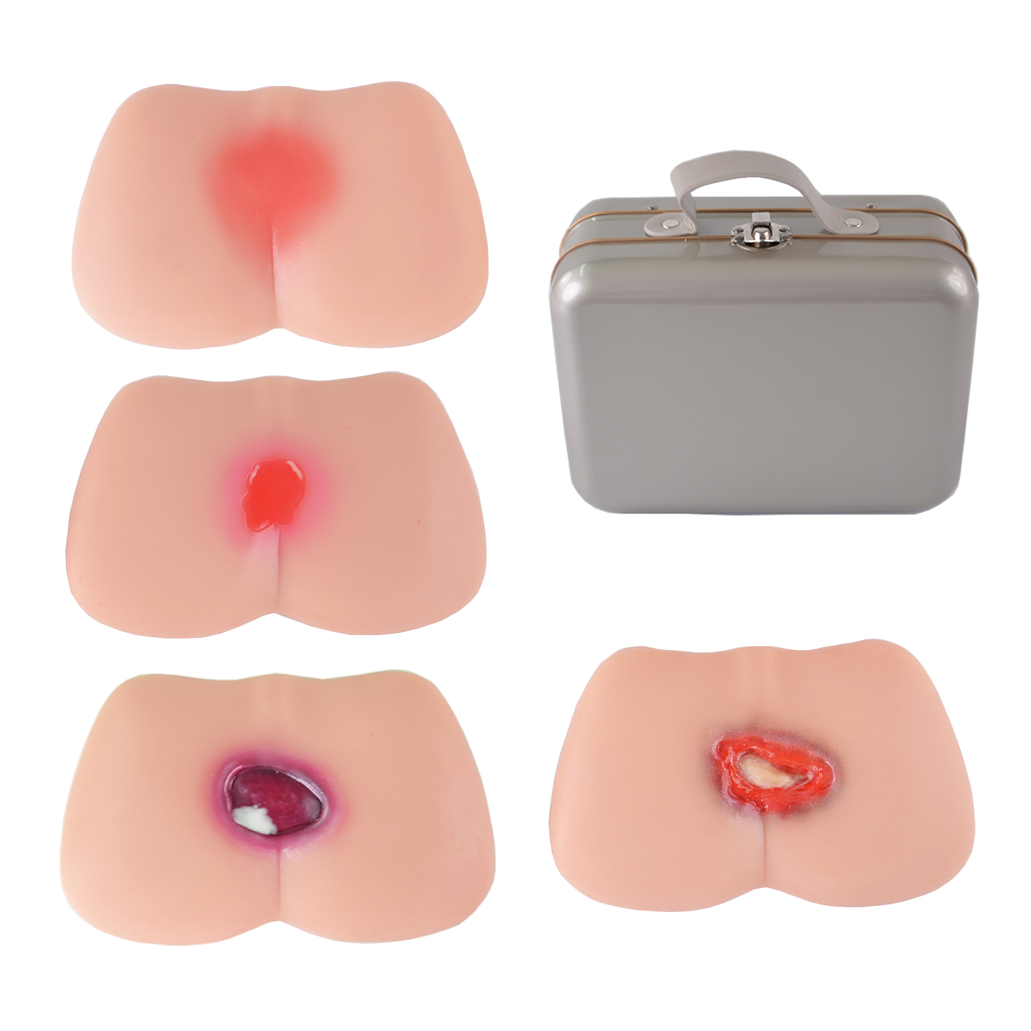
Telling a Stage 2 from an early Stage 3 — or spotting a deep tissue injury under intact skin — is a skill built through repeated, hands-on exposure. Training with a realistic staged pressure ulcer model lets nursing students and staff practise assessment, staging, and dressing selection on lifelike wounds before they care for real patients. The clearer a clinician’s mental picture of each stage, the earlier they intervene — and earlier intervention is what keeps pressure injuries from progressing.
For nurses, pressure ulcer care comes down to two disciplines: accurate staging — including avoiding the classic pitfalls — and matching the right treatment to each stage. Master both, and you intervene earlier, document defensibly, and stop most injuries before they ever become severe.
References & Further Reading
- National Pressure Injury Advisory Panel (NPIAP) (npiap.com)
- European Pressure Ulcer Advisory Panel (EPUAP) — International Pressure Ulcer Guideline (epuap.org)
- Wound, Ostomy and Continence Nurses Society (WOCN) (wocn.org)
- Wounds International — Pressure Injury Management (woundsinternational.com)
- Merck Manual — Pressure Injuries (merckmanuals.com)
- StatPearls / NCBI Bookshelf — Pressure Injury (ncbi.nlm.nih.gov/books)
Disclaimer: This article is for educational purposes only and is not a substitute for professional medical advice, diagnosis, or treatment. Always consult a qualified healthcare professional. Last updated: June 2026.