Dog extubation is not simply the last step of anesthesia. It is a planned transfer from a protected airway to a patient that must breathe effectively, keep its upper airway open, and protect itself from saliva, blood, or regurgitated material.
The safest timing varies with the dog, the procedure, the anesthetic protocol, body temperature, airway anatomy, and recovery quality. A single swallow or a fixed number of minutes should not replace a complete readiness assessment.
This guide explains how veterinary teams can prepare for extubation, evaluate a dog’s airway reflexes and ventilation, remove the endotracheal tube in a controlled way, and recognize complications that require immediate support or reintubation.
For tube selection, positioning, laryngeal landmarks, and placement confirmation before recovery, see How to Intubate a Dog: A Step-by-Step Veterinary Guide. For tube size, depth, cuff sealing, and placement troubleshooting during anesthesia, see Canine Endotracheal Tube Placement.
Why the Extubation Phase Deserves Its Own Plan
The endotracheal tube protects the airway only while it remains correctly positioned and connected to appropriate support. After it is removed, residual anesthetic drugs, hypothermia, pain, upper-airway swelling, secretions, regurgitation, or abnormal airway anatomy can still compromise breathing.
AAHA reports that a large proportion of anesthetic-related deaths in dogs and cats occur during the postoperative period, with many occurring within the first three hours. Recovery therefore requires the same deliberate monitoring and staffing as anesthetic maintenance.
Build the plan around one question: how much reserve does this dog have? Plan for a later, more controlled extubation in patients with:
- airway anatomy or disease — brachycephalic conformation, laryngeal paralysis or swelling, a neck mass, or previous airway surgery;
- surgery near the airway — oral, dental, pharyngeal, nasal, or upper-airway procedures;
- aspiration risk — regurgitation, vomiting, or megaesophagus;
- limited respiratory reserve — obesity, sleep-disordered breathing, pulmonary disease, hypoxemia, or recent respiratory distress; or
- a demanding anesthetic — hypothermia, prolonged recovery, major trauma, emergency surgery, significant blood loss, or previously difficult intubation.
Brachycephalic dogs deserve particular attention: their narrow, load-sensitive upper airways can destabilize as anesthetic depth changes, and apparent arousal does not prove that pharyngeal tone has returned. Keep them intubated until they are clearly ready, minimize agitation and heat stress, and have a reintubation plan immediately available.
With that risk established, agree the rest before recovery begins: who assesses readiness and removes the tube, which physiologic targets must be met, how the dog will be positioned, where oxygen, suction, a laryngoscope, and replacement tubes are kept, and what findings will trigger escalation or reintubation.
Equipment to Prepare Before Extubating a Dog
Do not dismantle the airway setup as soon as the procedure ends. Keep the following within reach until the patient is breathing safely after extubation:
- a functioning oxygen source and suitable delivery device;
- pulse oximetry and continued physiologic monitoring;
- capnography while the endotracheal tube remains in place;
- suction with an appropriate catheter;
- a laryngoscope with a checked light;
- the previous tube size plus smaller replacement tubes;
- a syringe for cuff deflation;
- materials to maintain head and neck position;
- emergency drugs and ventilation equipment; and
- an IV catheter that remains patent.
AAHA recommends leaving the IV catheter in place until the patient has been extubated, is in sternal recumbency, and has physiologic parameters returned to an acceptable range. This preserves rapid access if analgesia, sedation, fluids, or emergency drugs are required. For a refresher on placement and patency, see the canine IV catheterization guide.
When to Extubate a Dog: Five Readiness Checks
Extubation readiness is a pattern of findings, not a stopwatch event. Before removing the tube, assess ventilation, oxygenation, airway protection, recovery depth, and procedure-specific risks.
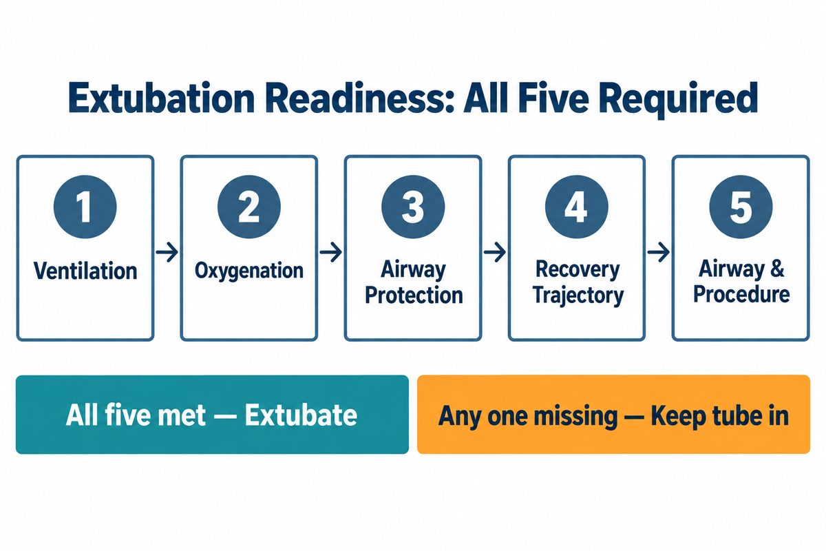
1. Spontaneous ventilation is adequate
The dog should have a consistent respiratory pattern with appropriate chest excursion and airflow. Respiratory rate alone can mislead: rapid shallow breaths may provide poor ventilation, while a slower pattern may be acceptable if tidal volume and gas exchange are adequate.
Continue capnography while the tube is in place when available. A rising end-tidal carbon dioxide level, decreasing respiratory effort, or apnea indicates that the dog may not be ready to maintain ventilation without support.
2. Oxygenation is acceptable
Pulse oximetry should be stable and interpreted together with oxygen delivery, perfusion, respiratory effort, and waveform quality. A normal saturation while the dog is still receiving supplemental oxygen does not prove that it will remain normal after the airway device is removed.
AAHA recommends that respiratory rate and SpO2 be within acceptable limits before extubation and that pulse oximetry continue through recovery.
3. The dog can protect its airway
A vigorous swallow is a useful sign that protective reflexes are returning, but it should not be used in isolation. Also assess purposeful tongue and jaw movement, improving head control, response to the tube, handling of secretions, and the ability to maintain a patent upper airway.
A weak, isolated swallow in a deeply sedated or hypothermic dog is not the same as sustained airway protection.
4. Anesthetic recovery is progressing appropriately
Consider the drugs used, their duration, whether reversal agents were administered, anesthetic length, pain control, and body temperature. Hypothermia can prolong drug effects and delay a coordinated recovery.
The dog does not have to be standing before extubation, but it should be recovering in a predictable direction rather than becoming progressively less responsive.
5. The airway and procedure are ready
Inspect and suction the mouth or pharynx when indicated. Confirm that throat packs have been removed and documented. Reassess bleeding, swelling, secretions, and regurgitation risk after dental, nasal, oral, or airway procedures.
How to Extubate a Dog Step by Step
1. Assign one person to the airway
One trained team member should control the tube and focus on the dog while another person assists with monitoring, suction, positioning, or equipment. Avoid making extubation an unannounced action during patient transfer.
2. Position the dog and breathing circuit safely
Choose a position that supports ventilation and allows secretions to drain without twisting the tube. Many higher-risk airway patients benefit from sternal recumbency with the head and neck supported in a neutral, open position.
Disconnect the breathing circuit before turning or moving the dog so the circuit does not torque, advance, or partially withdraw the tube. Reconnect oxygen and monitoring after repositioning if the tube is to remain in place.
3. Clear material that could threaten the airway
Inspect the oral cavity and suction visible saliva, blood, irrigation fluid, or debris when clinically indicated. Confirm again that no gauze or throat pack remains.
Do not perform aggressive blind suctioning. It can traumatize tissue, stimulate coughing, or push material deeper.
4. Perform the final readiness check
Immediately before extubation, confirm:
- stable spontaneous breathing;
- acceptable oxygenation;
- active airway-protective responses;
- appropriate recovery progression;
- no unresolved tube obstruction or circuit problem; and
- rescue equipment and personnel ready.
If the findings are mixed, keep the airway protected while the team corrects reversible problems such as hypothermia, excessive anesthetic depth, retained secretions, pain, or poor positioning.
5. Manage the cuff according to the procedure
For routine extubation, AAHA advises fully deflating the cuff immediately before removing the endotracheal tube. Verify deflation rather than assuming that air has been removed because the pilot balloon feels soft.
Dental and rhinoscopy patients are a specific exception described by AAHA. Positioning the nose slightly lower than the back of the head and leaving the cuff slightly inflated during withdrawal may help move blood or debris out of the trachea and into the pharynx, where it can drain from the mouth or be swallowed. This technique is procedure-specific and should not become the default for every dog.
6. Remove the tube smoothly
Untie the tube, stabilize the head and neck, and withdraw it in one controlled motion along the natural airway path. Avoid twisting the dog’s head, pulling against a partially inflated cuff in routine cases, or allowing the patient to drag the tube against its teeth.
Have suction and oxygen ready before the tube leaves the mouth. The person removing the tube should continue watching the laryngeal and respiratory response rather than immediately turning away to clean equipment.
7. Reassess immediately
Within seconds of extubation, evaluate:
- airway noise and patency;
- respiratory rate, pattern, and effort;
- chest excursion and airflow;
- mucous membrane color;
- SpO2 trend and signal quality;
- head and neck position;
- alertness and response; and
- coughing, gagging, regurgitation, or oral fluid.
Document the extubation time, readiness findings, cuff method, oxygen support, patient position, and any complications or interventions.
Monitoring After Dog Extubation
An apparently smooth tube removal does not end the recovery period. Residual respiratory depression can cause hypercapnia and hypoxemia, while swelling, soft-tissue relaxation, or secretions can progressively narrow the upper airway.
Continue monitoring:
- respiratory rate, pattern, effort, and airway sounds;
- SpO2 and oxygen requirement;
- heart rate and rhythm;
- blood pressure when indicated;
- body temperature;
- pain, dysphoria, and sedation;
- ability to maintain sternal recumbency and normal head position; and
- signs of regurgitation or aspiration.
The dog should be observed until alert, warm enough, physiologically stable, and ambulatory unless it could not walk before anesthesia. Monitoring frequency should reflect patient risk; a brachycephalic dog after airway surgery needs a different recovery plan from a healthy dog after a brief uncomplicated procedure.
Do not confuse agitation with adequate recovery. Pain, dysphoria, hypoxemia, hypercapnia, bladder discomfort, or fear can all cause restlessness. Reassess the patient rather than assuming that an active dog has a safe airway.
Common Complications After Extubation
Upper-airway obstruction
Soft palate obstruction, laryngeal dysfunction, swelling, blood, mucus, or abnormal neck position can restrict airflow. Warning signs include stridor, stertor, paradoxical chest movement, marked inspiratory effort, anxiety, cyanosis, and a falling SpO2.
Reposition the head and neck, provide oxygen, inspect the airway, and escalate promptly. Do not wait for complete obstruction before preparing to reintubate.
Hypoventilation or apnea
Residual anesthetic or opioid effects, hypothermia, neurologic disease, or respiratory muscle fatigue can reduce ventilation. A dog may appear quiet while carbon dioxide rises.
Stimulate and support the patient according to protocol, reassess drug effects and temperature, and be prepared to provide assisted ventilation and replace the airway.
Hypoxemia
Hypoxemia may result from hypoventilation, upper-airway obstruction, atelectasis, pulmonary disease, aspiration, or ventilation-perfusion mismatch. Check the pulse-oximeter signal, but treat the patient rather than dismissing a persistent low value as equipment error.
Regurgitation and aspiration
Regurgitated material can enter the airway before, during, or after extubation. Lower the head appropriately when safe, suction visible material, provide oxygen, and follow the veterinarian’s aspiration-response protocol. New coughing, tachypnea, increased effort, fever, or hypoxemia after a suspected event requires evaluation.
Coughing and airway irritation
Brief coughing can occur as the tube is removed. Persistent, forceful, or worsening coughing may reflect laryngeal irritation, secretions, aspiration, tube trauma, or airway disease.
Delayed or rough recovery
Hypothermia, pain, drug accumulation, metabolic abnormalities, hypoxemia, and hypercapnia can delay recovery. Thrashing or vocalization may indicate dysphoria, pain, fear, or physiologic compromise. Protect the dog from injury while the team identifies and treats the cause.
When Should a Dog Be Reintubated?
Reintubation is not a failure; it is a rescue action when the dog cannot maintain a safe airway or adequate gas exchange.
Prepare to reintubate when there is:
- apnea or ineffective ventilation;
- severe or progressive upper-airway obstruction;
- falling SpO2 despite appropriate oxygen and initial support;
- cyanosis or deteriorating consciousness;
- inability to manage secretions or protect the airway;
- persistent regurgitation with high aspiration risk;
- severe respiratory fatigue; or
- any rapidly worsening airway problem that cannot be corrected immediately.
Call for help early. Reintubation becomes harder after swelling, repeated attempts, hypoxemia, or patient agitation develops. Use the difficult-airway plan and the most experienced available operator.
Conclusion
Safe dog extubation depends on readiness, preparation, and continued observation. The team should confirm effective spontaneous ventilation, acceptable oxygenation, active airway protection, appropriate recovery depth, and a clear rescue plan before removing the tube.
The highest-risk period does not end when the endotracheal tube leaves the mouth. Careful positioning, continuous respiratory assessment, temperature and pain management, and early response to obstruction or hypoventilation are essential until the dog is alert and physiologically stable.
Frequently Asked Questions
Is one swallow enough to know a dog is ready for extubation?
No. A vigorous swallow is useful evidence that airway reflexes are returning, but readiness should also include adequate spontaneous ventilation, acceptable SpO2, purposeful recovery, secretion control, and the ability to maintain a patent upper airway.
Why might a dog cough after the breathing tube is removed?
Brief coughing can result from normal airway stimulation during tube removal. Persistent or worsening coughing may indicate irritation, secretions, aspiration, trauma, or underlying airway disease and should be reassessed.
How long should a dog be monitored after extubation?
There is no single duration for every patient. AAHA notes that many anesthetic-related deaths occur during the postoperative period, often within the first three hours. Monitoring should continue until the dog is alert, normothermic, physiologically stable, and ambulatory unless it was nonambulatory before anesthesia, with longer or more intensive observation for high-risk patients.
Sources and Veterinary Disclaimer
- AAHA: 2020 Anesthesia and Monitoring Guidelines for Dogs and Cats
- AAHA: Anesthetic Protocols and Recovery From Anesthesia
- AAHA: Troubleshooting Anesthetic Complications
- Gruenheid et al.: Risk of Anesthesia-Related Complications in Brachycephalic Dogs
- Vicenti et al.: Respiratory Effects of CPAP During Recovery in Brachycephalic Dogs
- Costa et al.: Postoperative Regurgitation and Respiratory Complications in Brachycephalic Dogs
- Johnson et al.: Isoflurane vs Sevoflurane Induction and Recovery in Adult Dogs
- Nagahama: Timing-Controlled Extubation Concept for Brachycephalic Dogs — a hypothesis-generating framework, not a validated routine protocol.
This article is for veterinary education and supervised simulation only. It does not replace a veterinarian’s assessment, formal anesthesia training, the endotracheal tube manufacturer’s instructions, or an institution’s recovery and emergency protocols.
Training Resources
Extubation competency belongs to the recovery phase, not to induction. Rehearse readiness assessment, cuff deflation, controlled tube removal, and the first minutes of unassisted breathing on a species-appropriate canine airway simulator, and run the escalation sequence — oxygen, suction, laryngoscope, replacement tube — until it is automatic. Human airway models represent different anatomy and should not be used as a substitute for canine-specific competency training.