Cat vs Dog Venipuncture: What Actually Changes
Four venipuncture defaults change when you move from dogs to cats: which hind limb vein you use, where the jugular ranks, how much volume margin you have, and how far patient tolerance drives the plan.
CONTACT EMAIL: [email protected]

Contact: [email protected]
Four venipuncture defaults change when you move from dogs to cats: which hind limb vein you use, where the jugular ranks, how much volume margin you have, and how far patient tolerance drives the plan.
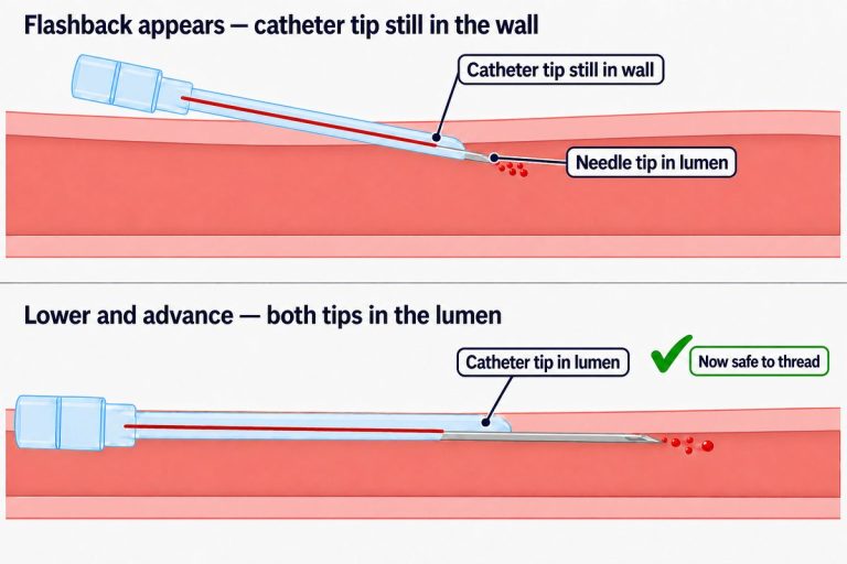
Place a peripheral IV catheter in a cat’s cephalic vein: setup, what differs from a dog, the insertion sequence, faint flashback, and why attempts fail.
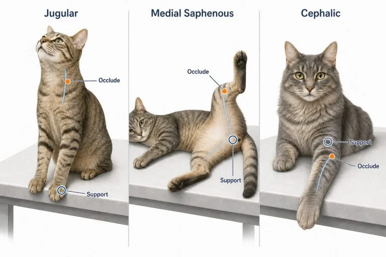
How to hold and position a cat so a blood draw or IV placement succeeds: what to prepare, positioning for each vein, when a towel helps, the signals that mean stop, and how to split the job between two people.
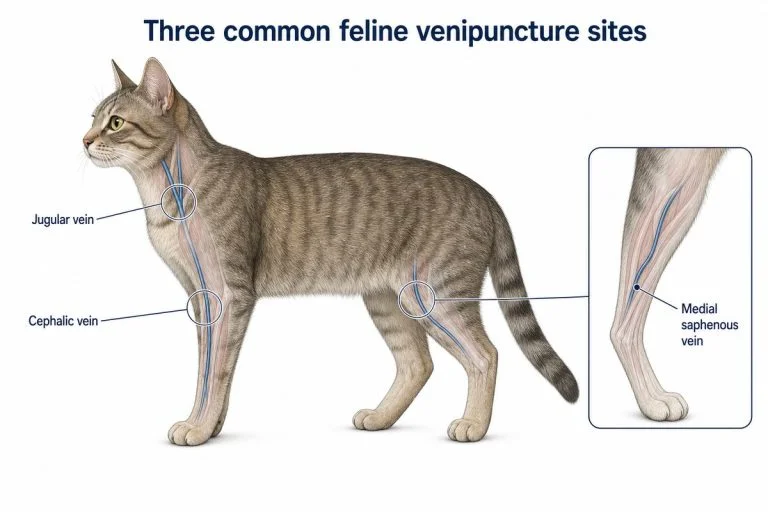
Choose a feline blood collection site by sample volume, patient tolerance, and the need to preserve a vein for IV access. Compares the jugular, medial saphenous, and cephalic veins in cats.
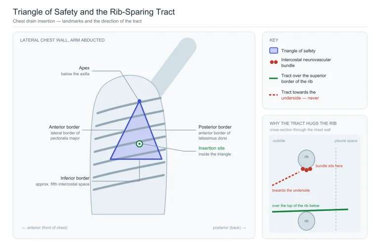
A chest drain insertion places a tube through the chest wall into the pleural space to evacuate air, blood, or fluid. Safety is decided before the blade touches skin: the correct side, a site inside the triangle of safety, and a tract passing over the top of a rib rather than under it. This article…
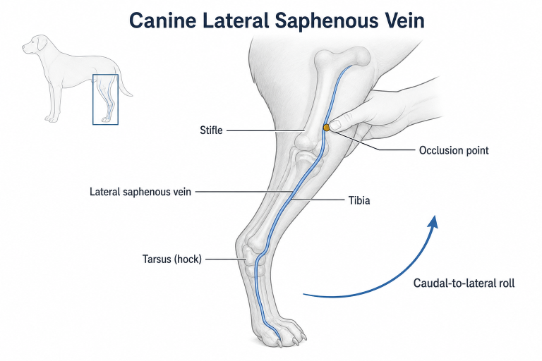
Learn canine lateral saphenous vein landmarks, hindlimb positioning, occlusion, stabilization, blood collection, and troubleshooting.
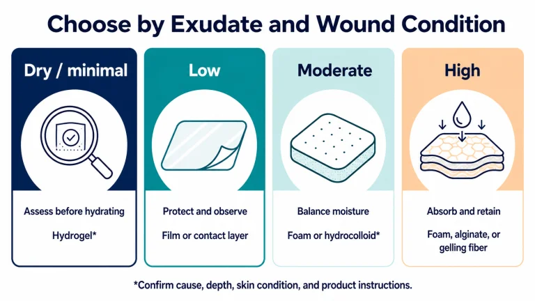
Compare wound dressing types by exudate, depth, wound condition, and skin tolerance, including film, foam, hydrogel, alginate, and contact layers.
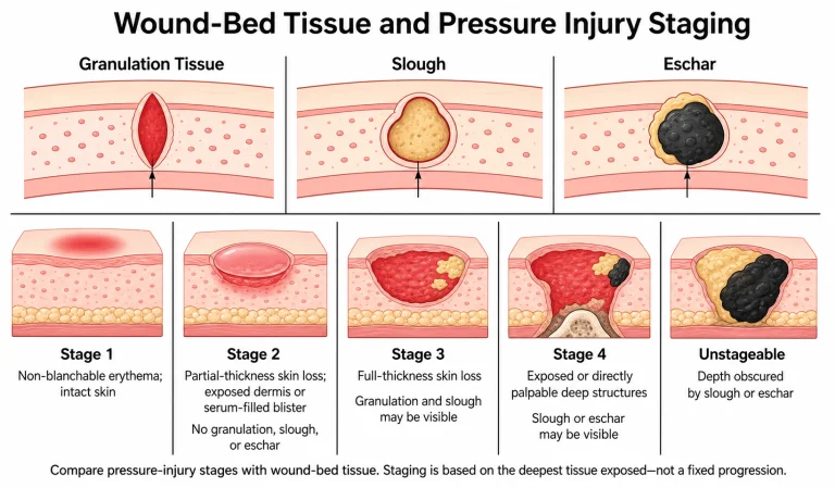
Granulation tissue is the new, vascular tissue that fills an open wound as it heals. Healthy granulation is usually a welcome sign, but color alone cannot tell you whether a wound is progressing normally. Red tissue may be healthy granulation, fragile hypergranulation, inflammation, or another abnormal change; yellow or black tissue may represent slough or…
A useful wound packing scenario changes more than the shape of the wound. It gives the learner a different access problem, decision, failure signal, or reassessment challenge. This guide shows instructors how to build five progressive stations—from a visible laceration cavity to a mixed scenario—without making the exercise unnecessarily complicated. Three Principles for Every Station…
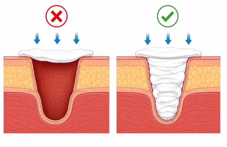
A wound can look packed and still be uncontrolled. This guide helps instructors recognize ten distinct failure patterns—not ten variations of “the learner packed it wrong.” By the end, you will be able to separate a poor method choice from a missed source, a loose fill, a pressure break, and a failed reassessment, then give…
End of content
End of content
Don't have an account yet? Sign up
No items in your cart. Go on, fill it up with something you love!
Start Shopping Now